Human Skeletal and Muscular Systems
| ✅ Paper Type: Free Essay | ✅ Subject: Biology |
| ✅ Wordcount: 1802 words | ✅ Published: 25 May 2018 |
- Rumana Anam
Describe the Human Skeletal and Muscular System in detail

(legacy, 2015)
Human Skeletal System
There are two types of skeletal system:-
Axial Skeleton
Axial Skeleton, this consists of skull, cranium and facial bones. Hyoid bone which includes tongue and muscles, which help to swallow. Vertebral column includes vertebrae and disks. Bony thorax includes ribs and sternum.

(Creationwiki, 2014)
Appendicular Skeleton
Appendicular skeleton, consists of pectoral gridle which include clavicle and scapula. Upper limbs which are the arms. Pelvic gridle which include sacrum and coccyx. Lower limbs which include the legs.

(Teachpe, 2015)
The properties and functions of tendons, ligaments and cartilages are as followes:-
Tendons
Tendons connect muscles to bones. Tendons let muscles move the skeleton, and are not very elastic. Tendons can be torn if too much pressure is put upon them.

(Fall down 7 times get up 8, 2012)
Ligaments
Ligaments connect bone to bone they hold together joints, they are tough elastic fibres. Ligaments stop bones from moving out of position when undergoing physical activity. If ligaments are pulled or twisted too far ligaments can tear and joints can be dislocated.

(Health-advisors, 2015)
Cartilages
Cartilages stop the ends of bones from rubbing together at the joints. It has a slippery surface which lubricate the joints.

(Orthopediatrics, 2012)
Joints
Where two or more bones meet together this is called as a joint. Without joints the human body would not be able to move. Joints, muscles and the skeleton system, are responsible for a huge range of movements that the human body is able to move (Auther Stream, 2014).
There are three different joint types:-
Fibrous Joints
Fibrous joints are fixed immovable joints, it is held together by dense irregular connective tissues. This joint allows no movement at all, and these joints are found in the skull and teeth.

(Redzuannorazlan, 2015)
Cartilaginous Joints
Cartilaginous joints are slightly movable joints, it allow little or no movement. Cartilaginous joints are tightly connected by cartilage. These joints can be found in pubic symphsis.

(Cnx, 2015)
Synovial Joints
Synovial joints are movable joint’s, it allows joints to move freely. Ligaments hold a synovial joint together. Smooth covering of cartilages stop bones from rubbing together and provide some shock absorption. Synovial joints consist of the following:-
Articular Capule – it is a sleeve like capsule that encloses and protects the synovial cavity. It is composed of two layers, 1st layer an outer fibrous capsule. 2nd layer an inner synovial membrane.
Synovial Fluid – lines the capsule and secrets synovial fluids. The function of the synovial fluid is to lubricate the joint, absorb shocks, supply oxygen and nutrients to the cartilage and to remove carbon dioxide and metabolic waste from the cartilage.
Bursae – it is a sac like structure containing a similar fluid to synovial fluid. It is located between tendons, ligaments and bones, it cushions the movement of these body parts.
Tendons Sheaths- it wraps around tendons to reduce frictions at the joints.

(Wisegeek, 2015)
Types of synovial joints
There are six types of synovial joints
- Planer – planer joints bones have two flat faces of bones sliding past each other, they allow a little movement in all direction. This joint is found in Mid-Carpal and Mid-Tarsal.
- Hinge – hinge joints allows extension and retraction i.e. forward and backwards movement. This joint can be found in the elbows and knees.
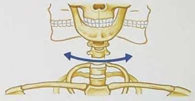
- Pivot – A ring of bone that fits over a bone protrusion, around which it can rotate, this is called as a pivot joint. This joint only allows rotation and can be found between the atlas and the axis in the neck (Auther Stream, 2014).
- Condyloid – condyloid joints have an oval shaped bone which fits onto a corresponding shaped bone. The joint allows forwards, backwards, left and right movement. This joint can be found between the metacarpals and phalanges in the hand (Auther Stream, 2014). .
- Saddle – saddle joints have a cancave and canvex shaped bones, which allow forward, backwards, left and right movement. This joint can be found in the thumb.
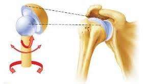
- Ball and Socket – ball and socket joint has a round shaped bone which fit into a cup shaped bone. This joint allows movement in all direction including rotation, most mobile joints are ball and socket i.e. the shoulders and hips.
Planer Joint

(Studyblue, 2014)
Hinge Joint

(Cae2k, 2001)
Pivot Joint
(Yiss Anatomy, 2010-11)
Condyloid Joint

(Yiss Anatomy, 2010-11)
Saddle Joint

(Studyblue, 2014)
Ball and Socket Joint
(Pivotalphysio, 2014)
B – Describe and compare the properties of different types of muscles, also discuss the sliding filament hypothesis of muscle contraction.
Muscle Tissues
Muscle cells are highly specialised for contractions.The muscle contractions can produce movement of the whole body and or a part the body, providing that the muscles are attached to a movable part of the skeleton. Within the wall of a hollow organ, if there is muscle there, the muscle contractions may allow the contents of the organ to move, e.g. peristaltic movement of material in the digestive tract. There are three types of muscle tissues (Kentsimmons, 2015).
1. Smooth Muscle Tissue
Smooth muscle can be found throughout the internal organs of the body especially in areas such as the digestive tract. As its contraction isnotunder conscious nervous control, it is referred to asinvoluntarymuscle.
Smooth muscle fibres arespindle-shapedstructures with aprominent centrally located nucleus.They are shorter in length and theydo not exhibit striations. The cells happen when the individual fibres within the organs or as groups of fibres tightly interweave in sheets or bands (Kentsimmons, 2015).

(Legacy, 2015)
2. Skeletal Muscle Tissue
Skeletal muscles form the flesh of the body. They are attached to the motion of the bones of the skeleton. For example, the biceps, brachii and pectoralis are skeletal muscles as they are attached to the motion of the bone. As the contraction of the skeletal muscles is underconsciouscontrol, they are also called voluntary muscles.
Skeletal muscle cell are highly customised, large,multi-nucleate cell(fibre). Each fibre iscylindrical in shapewithblunt, rounded ends. The flattenednucleiare located mainly at theperiphery of the cell, just inside the sarcolemma. The cross-striped (orstriated) seem of light and dark banding, which are the result from the arrangement of myofibrils, and small protein contractile units rooted in the sarcoplasm (Kentsimmons, 2015).

(Studyblue, 2014)
3. Cardiac Muscle Tissue
Cardiac muscle is a highly specialised tissuerestricted to the wall of the heart. It is aninvoluntary muscle, as its contractionis not controlled by the organism.
Cardiac fibres usually form long chains of cells thatbranch and intertwine this is different to smooth and striated fibres.The peculiar wringing action of the heart is a result of this assembly. The junction of one cell with another cell in aparticular chain is known as anintercalated discand looks like as a heavy dark line running across the fibre (Kentsimmons, 2015).
 (UCL, 2015)
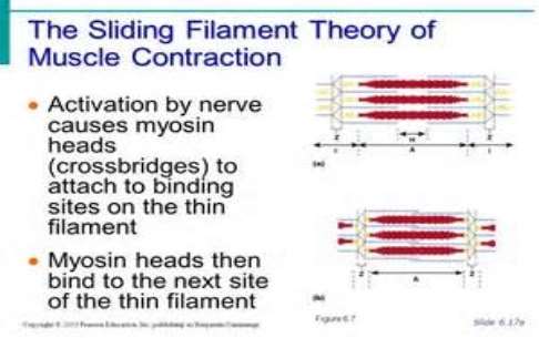
Sliding Filament Hypothesis
Muscle Contraction
Smaller fibre called myofibrils make up each muscle fiber. These contain even smaller units called actin and myosin filaments. These filament slide in and out between each other for muscle contraction. Sarcomeres are the smallest unit of skeletal muscle that contract, the repeat themselves over the length of the myofibrils.
Five steps of muscle contraction
There are five steps to muscle contraction
- A nervous impulse arrives at the central nervous system, which release a chemical called acetylcholine. The acetylcholine defuses the motor end plate which travels to the muscles by transverse tubules, causing calcium to be released.
- As there is a high concentration of calcium, calcium now binds to troponin changing its shape, so moving tropomyosin from the active state of actin. The myosin filament are now able to connect to the actin to form a cross bridge.
- The breakdown of ATP release energy which lets myosin to pull the actin inwards therefore shortening the muscle, this process happens throughout the length of every myofibril in the muscle cell.
- The myosin lets itself go from actin and the cross bridge is now broken, when the ATP molecules bind to the myosins head. When ATP is broken down the myosin heads can be again attach to actin binging further actin filaments and repeating the process.
- The process of muscular contraction can last as long as there are enough ATP and calcium stores. Once the impulse is stopped the calcium is pumped back and actin returns to its resting place (Teach PE, 2015).
(Slideplayer, 2011)

(Human Biology Lab, 2014)
Stretched muscles have I band and H zones is elongated due to reduced overlapping of actin and myosin filaments.
Partially concentrated muscles have more overlapping actin and myosin filaments with potential for cross bridges to form i.e. where I band and H zones are shortened.
Fully contracted muscles have most actin and myosin filaments overlapping. As the thin layer have overlapped there are reduced potential for cross bridges to form, therefore there will be a low force production from the muscle.
References
Teach PE 2015, Sliding Filament Theory – Skeleton and Muscle, [online] available at http://www.teachpe.com/anatomy/sliding_filament.php, Last accessed 10/04/15
Kentsimmons 2015, Muscle Tissue, [online] available at http://kentsimmons.uwinnipeg.ca/cm1504/15lab42006/lb4pg7.htm, Last accessed 10/04/15
Auther Steam2014, Joints, [online] available at http://www.authorstream.com/Presentation/anu_jinnee002-656673-joints/, Last accessed 10/04/15
Legacy 2015, Language of Anatomy, [online] available at legacy.owensboro.kctcs.edu/gcaplan/anat/Notes/API%20NOtes%20A%20Body%20Systems%20and%20Cavities.htm, Last accessed 10/04/15
Creation Wiki 2014, Axial Skeleton, [online] available at http://creationwiki.org/Axial_skeleton, Last accessed 10/04/15
Fall down 7 times get up 8 2012, A is for Achilles, [online] available at http://falldown7timesgetup8justdontgiveup.blogspot.co.uk/2012/01/is-for-achilles.html, Last accessed 10/04/15
Health Advisor 2015, Ulnar Collateral Ligament, [online] available at http://health-advisors.org/ulnar-collateral-ligament/, Last accessed 10/04/15
Orthopediatrics 2012, Anatomy, [online] available at http://www.orthopediatrics.com/docs/Guides/OCD_elbow.html, Last accessed 10/04/15
Redzuannorazlan 2015, Osteologi, [online] available at http://redzuannorazlan.blogspot.co.uk/2010/10/osteology-terms.html, Last accessed 10/04/15
CNX 2015, Cartilagious Joint, [online] available at http://cnx.org/contents/98ad6c26-7214-4875-af25-6c108fc54b92@3/Cartilaginous_Joints, Last accessed 10/04/15
Wise Geek 2015, What is a joint capsule, [online] available at http://www.wisegeek.org/what-is-a-joint-capsule.htm, Last accessed 10/04/15
Studyblue 2014, Joints, [online] available at https://www.studyblue.com/notes/note/n/chapter-8/deck/4718069, Last accessed 10/04/15
Cae2k 2001, Hinge Joints, [online] available at http://cae2k.com/how-to-send-photos-to-your-phone-0/hinge-joints.html, Last accessed 10/04/15
Yiss Anatomy 2010-11, Joints and Movements, [online] available at http://yiss-anatomy2010-11.wikispaces.com/Anne+and+Yeji, Last accessed 10/04/15
Pivotal Physio 2014, Pivotal Physiotherapy, [online] available at http://pivotalphysio.com/?p=1597, Last accessed 10/04/15
Legacy 2015, Muscle Types, [online] available at legacy.owensboro.kctcs.edu/gcaplan/anat/Notes/API%20NOtes%20A%20Body%20Systems%20and%20Cavities.htm, Last accessed 10/04/15
Studyblue 2014, Muscles, [online] available at https://www.studyblue.com/notes/note/n/chapter-8/deck/4718069, Last accessed 10/04/15
UCL 2015, Cardiac Muscle, [online] available at http://www.ucl.ac.uk/~sjjgsca/MuscleCardiac.html, Last accessed 10/04/15
Slideplayer 2011, The Muscular System, [online] available at
http://slideplayer.com/slide/772255/, Last accessed 10/04/15
Human Biology Lab 2014, How do Muscles Contract, [online] available at http://humanbiologylab.pbworks.com/w/page/70195477/How%20DO%20muscles%20contract, Last accessed 10/04/15
Cite This Work
To export a reference to this article please select a referencing stye below:
Related Services
View all


DMCA / Removal Request
If you are the original writer of this essay and no longer wish to have your work published on UKEssays.com then please click the following link to email our support team:
Request essay removal