Heart, Cardiac Cycle and the Circulatory System
| ✅ Paper Type: Free Essay | ✅ Subject: Biology |
| ✅ Wordcount: 3358 words | ✅ Published: 08 Jun 2018 |
(i) Explain the function of heart, and the structure of arteries, veins and capillaries.
The heart (left side) receives blood filled with oxygen (O2) which comes from the lungs. From there the blood is pumped throughout the body via the aorta and into blood vessels. On the right side is where the heart gets its deoxygenated blood (CO2) which in turn is then sent to the lungs for cleansing. (ivyroses.com 2016)
Arteries are made up of 3 layers. The tunica interna is the inner layer. This layer is encased in connective tissue and elastic fibers. After that the next layer up is the tunica media. This thick layer consists mainly of smooth muscle. This layer supports the vessel itself and aids blood flow regulation. The outside layer is called the tunica externa. This layer is made up of connective tissue and elastic fibers. It has the ability to change and become looser connective tissue around the outside of the vessel. (training.seer.cancer.gov ND)
Veins carry blood back to the heart. This blood is filled with CO2. The blood is then sent to the lungs for purification and then back to the heart to be sent around the body. The only veins which carry oxygen are the umbilical and pulmonary veins. Veins are like straws, they are small in structure. They have a dense outer layer which consists of connective tissue. Underneath that is the middle layer which is made up of smooth muscle and finally the middle layer which is made up of endothelial cells. (reference.com ND)
Capillaries are tiny blood vessels inside the bodily tissues that carry blood from the arteries to the veins. They are more common in tissues like muscle tissues than in connective tissues. (biology.about.com ND)
Arteries divide into arterioles. These arterioles branch off into the muscle where they reach the capillaries. A capillary bed is then formed. This is a network of capillaries which then carry blood to the veins. Capillaries also move gases in and out of themselves. These gases include oxygen and carbon dioxide. (teachpe.com ND)
Like the lungs, capillaries are responsible for the process of diffusion. Oxygen separates from haemoglobin (found in red blood cells) and passes through the walls of the capillaries into muscle cells where it associates itself with the Myoglobin. This is the muscle cells version of haemogloblin. The oxygen is then used in aerobic metabolism to supply the muscle with energy. (teachpe.com ND)
Cross section of an artery.
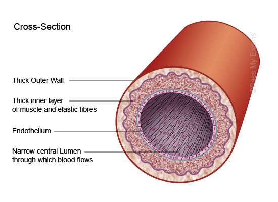
©Google Images
Cross section of a vein.
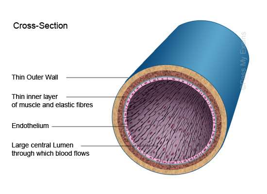
©Google Images
Cross section of a capillary.
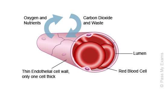
©Google Images
(ii) Explain the cardiac cycle and the flow of the blood through the main blood vessels by using the diagram.
Blood is pumped around the body in stages, namely the diastole stage and the systole stage. In the diastole stage the atria and ventricles are relaxed and allow blood to enter via the vena cava in to the upper right chamber (right atrium). The deoxygenated blood is passed through a valve which prevents the blood from running backwards, into the lower chamber (right ventricle). After this, the blood is then pumped under extreme pressure from the right ventricle into the lungs by the pulmonary artery. (quora.com 2017)
In the second stage oxygenated blood enters the left atrium. It then passes through a valve which closes over after the blood has passed through, preventing the said blood from running back into the atrium. Blood flows down into the left ventricle. It then goes to the aorta (main artery in the body) where it is pumped under immense pressure. Blood is pumped under high pressure to ensure it is sent to the rest of the body as it should do. (biology.about.com 2017)
(livescience.com 2017)
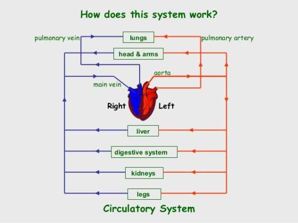
©Google Images
|
Arteries |
Veins |
Capillaries |
|
Contain narrow lumens |
Contain wider lumens |
Have one layer of cells. Here diffusion takes place. They are the smallest of all the blood vessels. |
|
Blood is under high pressure |
Blood is under low pressure |
|
|
Takes blood from the heart |
Takes blood to the heart |
Take blood form the body and exchange nutrients, O2 and waste with tissues. |
|
Have more muscle/elastic tissue |
Have less elastic tissue |
They are oozy vessels that serve as links between arterial and venous systems |
|
Carries O2 rich blood (except for the pulmonary artery) |
Carries CO2 rich blood (except for the pulmonary vein) |
|
|
Has no valves (except for the semi-lunar valves in the aorta and pulmonary artery |
Has valves in main veins – to stop waste materials returning to the tissues |
(iii) Explain the term blood pressure and describe the role it plays in circulatory system. Describe the condition high and low pressure.
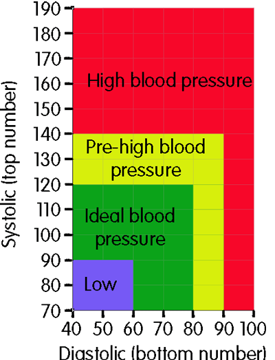
When the heart pumps blood around the body the blood pushes against the wall of the blood vessels. The rate at which it pushes against the walls is called blood pressure. If blood pressure is high your arteries are put under immense pressure to deliver blood around the body. This may result in the person suffering a heart attack or a stroke. Symptoms of high blood pressure include tiredness, irregular heartbeat and difficulty breathing. Low blood pressure if left untreated can be life threatening. A reading which is less than 90/60 is deemed to be low blood pressure. Although the causes are unclear, it is thought that dehydration and serious medical disorders are among the causes of low blood pressure. (mayoclinic.org ND)
What the numbers mean: A blood pressure reading is made up of two numbers, written as one over the other e.g. 80/120. The number at the top is known as the systolic blood pressure. This is the highest level that a person’s blood pressure will reach when the heart beats. The bottom number is called the diastolic blood pressure. This is the lowest level that a person’s blood pressure will reach when the heart relaxes between every single beat. (bloodpressureuk.org 2008)
 ©Google Images
Section B
Identify the main muscles groups within the body and interconnections between the muscular and skeletal system.
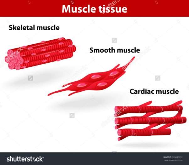
(ii) Differentiate between the three types of muscles, Smooth, Skeletal, and Cardiac.
|
Smooth |
Skeletal |
Cardiac |
|
Maintains flow of fluid in hollow structures |
Attached to the bone |
Only found in the heart |
|
Found in structures such as veins and arteries |
Relaxes and contracts |
Involuntary contraction |
|
Contract slowly |
Striated |
Features are endurance and consistency |
|
Arranged in bundles of muscle fibres. |
Contracts voluntarily |
Striated |
|
Have only one nucleus |
Soft and fragile |
Makes up the atria and ventricles |
|
Not banded |
Every fibre has lots of nuclei and is surrounded by a cover. |
Relaxes to fill the heart with blood |
|
Muscles work automatically |
Made up of cylindrical cells that made up fibres |
Never tires |
(Healdove.com 2016)
(Class notes 2016 S. Curran)
(training.seer.cancer.gov 2016)
©Google Images
(iii) Describe how the Skeletal and the Muscular System connect together to create the body movement.
Movement in the body happens when the skeletal and muscular systems work simultaneously. The skeleton gives us shape and without it we would just be one big blob of skin. The main function of these two systems is bodily movement. (Class Notes.S Curran.2016)
As well as joints, bones and muscles work hand in hand to form levers e.g. in the arm and the knee. (livestrong.com 2015) Tendons attach muscles to bones. This is allows bones and muscles to form such levers. (prezi.com 2010)
Bones have the ability to hoard fat in their cavities which in turn can help store minerals such as calcium and phosphorus. Calcium is a vital mineral because without it blood would be unable to clot and a lack of calcium would hinder bodily movement. (prezi.com 2010) Phosphorus enables the body to make protein which is necessary for growth and repairing damaged cells. (medlineplus.gov 2015)
Section C
Outline the composition of bone the structure of the long bone and explain the function of the skeleton
Identify and describe the composition of the bone, joint types and the function of the skeleton.
- Outline the composition of bone the structure of the long bone and explain the function of the skeleton.
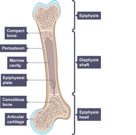
The bone is made up of 3 layers – periosteum, compact bone and spongey bone.
Periosteum is a double layered tissue that covers the compact bone. It cannot be seen by unaided vision. It’s an important layer because it holds cells called osteoblasts. These osteoblasts form new bones. The double layer also means that ligaments and tendons can anchor the bone.
(healthbase.wordpress.com 2016)
Compact bone accounts for 80% of the human skeleton. It forms around the spongey (cancellous) bone. The long bones of the body e.g. arm, leg etc. are primarily made up of compact bone.
(britannica.com 2016)
Cancellous bone is the spongey bone which is located at the end of each of the long bones and in the vertebrae of the spine. The cancellous bone contains pores as well as red bone marrow which is used to make red blood cells and stem cells which are then used to repair the bone if it gets damaged or broken. The spongey bone is much easier to fracture than the long bone because of its soft composition.
©Google Images
Functions of the skeleton:
Protection – the skeleton acts as a protector for major organs such as the heart and lungs which are protected by the rib cage and the breast bone.
Storage – bones store minerals such as calcium. If you consume too much of a particular mineral e.g. calcium, it can build up on the bone. Whenever the supply is low then the body makes up for it by drawing the excess minerals from the bone to build up the supply in the blood system.
Movement – bones along with muscles work together to make the body move.
Form blood cells – bone marrow makes red blood cells. On average 2.6 million cells are created per second.
(Class notes unit 3 2016)
Different types of joints and their functions.
There are 3 types of joints: Synovial, fibrous and cartilaginous.
Synovial joints are divided up in to 6 categories: Gliding joints, condyloid joints, saddle joints, hinge joints, ball and socket joints and pivot joints.
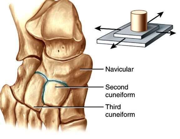
Gliding joints allow for smooth rotation in different directions along a smooth surface. An example of this type of joint is the carpal joint which is located in the wrist. (livestrong.com 2012)
©Google Images
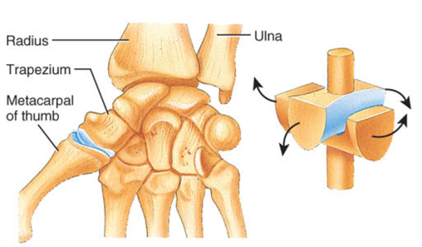
Saddle joints are so called because they fit like a rider on a saddle. They are able to bend in several different directions without ever actually sliding. A prime example of a saddle joint is the joint at the base of the thumb. (livestrong.com 2012)
©Google Images
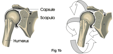
Ball and socket joint – This type of joint is usually found for example in the shoulder. As the name states a ball and socket joint is given that name because it is where one end of a long bone (ball) meets a socket. Ball and socket joints facilitate movement in several directions. (livestrong.com 2012)
©Google Images
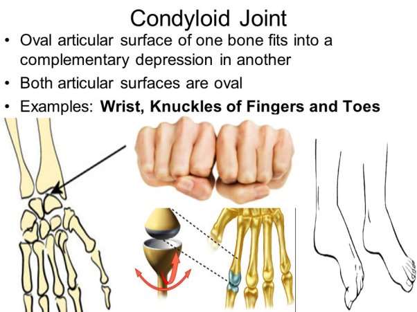
Condyloid Joints – Unlike gliding joints condyloid joints have not got a smooth surface. Bones rotate past each other. Condyloid joints can be found in the wrist. (livestrong.com 2012)
©Google Images
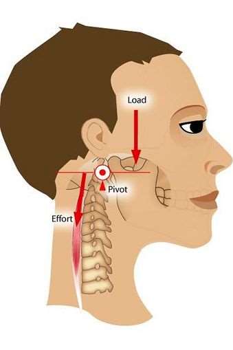
Pivot Joint – This type of joint moves but there is no gliding movement. It facilitates turning moves without any bending sideways as such. An example of a synovial joint is between the first and second vertebrae of the spine. It allows limited movement while keeping the head in place. (livestrong.com 2012)
©Google Images
Hinge Joints – These are formed between 2 bones. A hinge joint allows us to extend (elbow, knee etc.) freely, bones do not slide past each other. (livestrong.com 2012)
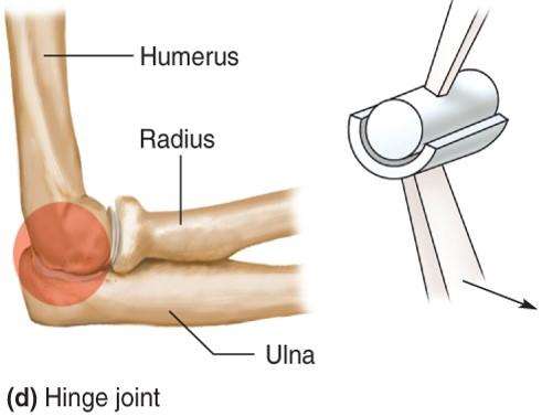
©Google Images
Fibrous Joints:
These are generally immovable joints. There are 3 types of fibrous joints – sutures, gomphoses and a syndesmosis.
Sutures are the joints which connect bones in the skull
Gomphoses are located between the jaw bone and the teeth.
A syndesmosis joint is where a ligament connects two bones for example in the leg the tibia and the fibula. It allows for a very slight movement.
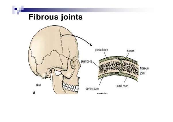
©Google Images
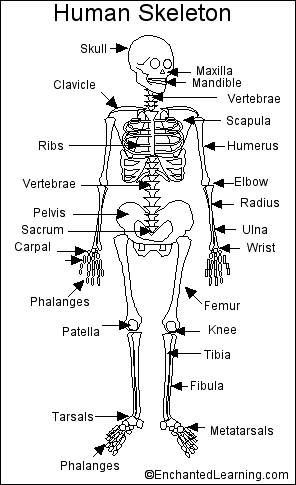
Label the main bone of appendicular axial skeletal. (see attached diagram)
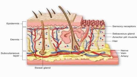
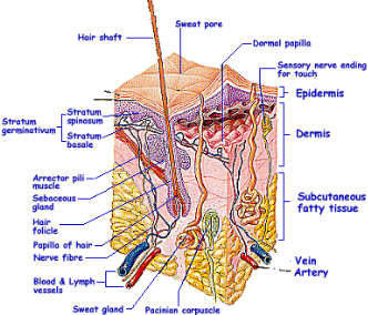
The main function of the skin, its structure and relationship between the skin, and circulatory and nervous system
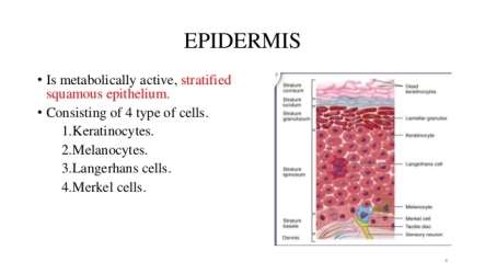
The Skin is one of the largest organs of the body. It is made up of three layers: The Epidermis, Dermis and the subcutaneous fatty tissue. As it has no blood vessels, the epidermis is dependent on the dermis to feed it nutrients and for disposing of excess waste.
Epidermis
 ©Google Images
Keratinocytes is the name given to the wall of the epidermis. It produces keratin, a protein that protects the epithelial cells. “The epidermis regenerates in orderly fashion by cell division of keratinocytes in the basal layer, with maturing daughter cells becoming increasingly keratinised as they move to the skin surface.” (Dermnetz.org 2016)
Keratinocytes are regenerated monthly. There are a number of cells within the epidermis:
Merkel cells – “Merkel cells are cells found in the basal layer of the epidermis.” (Dermnetnz.org 2016) These cells allow us to feel sensations such as pain, coolness, heat, numbness and to feel objects. These tiny cells can only be identified by using electron microscopy. They are most commonly found in the soles of the feet and the palm of the hand.
Dermis
©Google Images
This is the second layer of the skin and is the biggest part of the cross-section of the skin. In the dermis there are many things such as nerve endings, sweat glands and blood capillaries. The Dermis is sub-divided into 2 sections namely: Papillary Dermis and the Reticular Dermis. Collagen, Elastic Tissue and Reticular fibres are present throughout the Dermis.
Subcutaneous layer
 (c)Google Images
“The subcutaneous layer is an important line of defence, protecting the fragile organs and bones from outside forces, such as pathogens.”(reference.com 2016)
The Hypodermis or subcutaneous layer acts as a protector for organs and the skeleton against the elements. Like the other two layers, its thickness depends on where it is in the body. For example the subcutaneous layer on the eyelids would be thinner than on the skin around the soles of the feet. Its main function is to act as a temperature regulator.
Functions of the skin:
Protection: Melanin in the skin protects the body from harmful UV rays produced by the sun. Skin has the ability to protect organs and bones from exposure to the environment. It also can protect the body from bacterial infections.
Temperature Regulation: When environmental temperatures are high, the skin releases sweat as a means of cooling the body down. If you’re cold, blood vessels in your skin fill up with blood to generate heat.
Immunity – Some skin cells work with the immune system to fight against bacteria and viruses. Some bacteria live on the skin, this is normal as it is good bacteria. However if it gets in under the skin, it can cause infection such as cellulitis.
Growth and Movement– The skins elasticity allows the body to grow as we get older. Without it we would not be able to move freely, if at all.
Excretion – Skin helps to get rid of waste such as urea, sweat and carbon dioxide.
Endocrine: when exposed to a small amount of UV rays, the skin produces Vitamin D. a chemical in the skin called 7-dehydrocholesterol reacts with the UV rays. Over exposure to UV can have dire consequences such as skin cancer, so exposure to the sun must be kept in moderation.
Absorption: Skin absorbs oxygen and nitrogen. Some animals have no need for lungs because they can actually breathe through their skin.
Water Resistance: Skin is covered by oils and nutrients which form a protective layer against water. (newhealthguide.org ND)
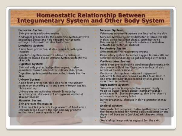
The relationship between the skin, circulatory system and the nervous system.
All bodily functions are regulated by the brain via the nervous system. The brain sends messages to the rest of the body through nerves. Without it the circulatory system could not function. Cardiac functioning and transportation of fluid in the body is started by messages coming from the brain. The circulatory system is made up of arteries, veins, capillaries and other passages which are central to its functioning. The nervous system is made up of the spinal cord, brain and nerves. It controls what the body does. Signals are sent to and from the brain via the nerves to create proper functioning. A prime example of how these systems work together is in blood pressure and regulation of the heart rate. The vagus nerve regulates the pumping of the heart. Blood is pumped through the passages around the body and into the organs. Baroreceptors give the brain information about blood pressure. The brain can then make adjustments to how the heart pumps. It is because of this process that blood pressure is kept at a normal level. The nervous system has the power of bodily functions but it needs the circulatory system to relay the messages so that such adjustments can be made accordingly. (wisegeekhealth.com ND)
The integumentary system (skin) works with the circulatory system. The skin contains networks of capillaries. This means that substances can enter the bloodstream though the skin and this is the reason why some medicines such as female contraception can be delivered in patch format. Neurons are contained within the skin to sense the environment. These neurons send signals to the nervous system such as touch and it begins action based on these signs. For example if you burn your finger, nerve cells send signals up your arm to the spinal cord and brain. Nerve cells in the brain interpret these messages as pain. Skin aids temperature regulation by way of changing blood supply patterns and by sweating which helps to cool the body down. (sciencenetlinks.com ND)
©Google Images
(iii) Using the table format give six examples of viral, bacterial, fungal skin diseases…..
|
Viral |
Bacterial |
Fungal |
|
Flu |
Tuberculosis |
Nail Fungus |
|
Shingles |
Cellulitis |
Oral Thrush |
|
Viral Pneumonia |
Salmonella |
Athlete’s Foot |
|
HIV/AIDS |
Helicobacter Pylori |
Impetigo |
|
Chicken Pox |
Staph Infection |
Jock Itch |
|
Herpes |
Scarlet Fever |
Vaginal Yeast Infection |
(Healthgrades.com 2016)
(Rightdiagnosis.com 2016)
(Healthonline.com 2016)
(Dermnetz.org 2016)
Example of a Viral Infection: 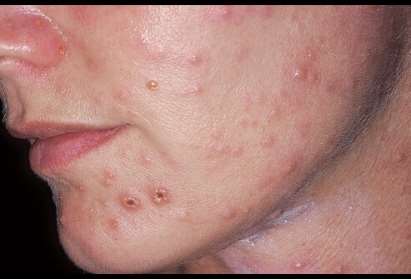 ©Google Images
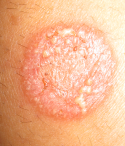
Example of a bacterial infection: 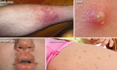 ©Google Images
Example of a fungal infection:
 ©Google Images
References:
(Class notes unit 3 S. Curran 2016)
Biology.about.com (2016) “Phases of the Cardiac Cycle” [online], available: http://biology.about.com/od/anatomy/ss/cardiac_cycle.htm[Accessed 1st March 2017]
Biology.about.com (2017) “What Is a Capillary?” [online], available:
Bloodpressureuk (2008) “What is blood pressure?” [online], available:http://www.bloodpressureuk.org/BloodPressureandyou/Thebasics/Bloodpressure [Accessed 15th March 2017]
Bloodpressureuk (ND) “What is blood pressure?” [online], available: http://www.bloodpressureuk.org/BloodPressureandyou/Thebasics/Bloodpressure [Accessed 19th January 2017]
Britannica.com (2016) “Compact bone” [online], available: https://www.britannica.com/science/compact-bone [accessed 12th December 2016]
Dermnetz.org (2016) “Bacterial Skin Infections” [online], available: http://www.dermnetnz.org/topics/bacterial-skin-infections/ [accessed 5th December 2016]
Healdove.com (2016) “The Differences Between Skeletal, Smooth & Cardiac Muscles” [online], available:https://healdove.com/misc/The-function-of-Muscles-and-the-3-main-types [Accessed 19th December 2016]
Healthbase.wordpress.com (2016) “Medical Tourism Blog, Surgical Tourism Blog, Overseas Medical Travel Blog” [online], available: https://healthbase.wordpress.com/2007/03/22/composition-of-a-bone/ [accessed 12th December 2016]
Healthgrades.com (2016) “What are viral diseases?” [Online], available: https://www.healthgrades.com/conditions/viral-diseases [accessed 5th December 2016]
Healthline.com (2016) “Candida Fungus Skin Infection” [online], available:
http://biology.about.com/od/anatomy/ss/capillary.htm [Accessed 2nd March 2017]
http://www.healthline.com/health/skin/candida-fungus#ReadThisNext0 [accessed 5th December 2016]
Ivyroses (2016) “The Functions of the Heart” [online], available: http://www.ivyroses.com/HumanBody/Blood/Heart_Functions.php [Accessed 18th January 2017]
Livescience (2016) “Circulatory System: Facts, Function & Diseases” [online], available: http://www.livescience.com/22486-circulatory-system.html [Accessed 1st March 2017]
Livestrong (2012) “6 types of synovial joints” [online], available: http://www.livestrong.com/article/74183-types-synovial-joints/ [Accessed 4th January 2017]
Livestrong (2012) “6 types of synovial joints” [online], available: http://www.livestrong.com/article/74183-types-synovial-joints/ [Accessed 5th January 2017]
Livestrong.com (2015) “How the Skeletal System Works With the Muscular” [online], available: http://www.livestrong.com/article/76374-skeletal-system-works-muscular/ [Accessed 4th January 2017]
Mayoclinic (ND) “Low blood pressure – Hypotension” [online], available: http://www.mayoclinic.org/diseases-conditions/low-blood-pressure/basics/definition/con-20032298 [Accessed 19th January 2017]
Medlineplus (2015) “Phosphorus in diet”[online], available: https://medlineplus.gov/ency/article/002424.htm [Accessed 4th January 2017]
Newhealthguide (ND) “Functions of the Skin” [online],
Cite This Work
To export a reference to this article please select a referencing stye below:
Related Services
View all


DMCA / Removal Request
If you are the original writer of this essay and no longer wish to have your work published on UKEssays.com then please click the following link to email our support team:
Request essay removal