Histological Features of Skeletal Muscle
| ✅ Paper Type: Free Essay | ✅ Subject: Physiology |
| ✅ Wordcount: 1288 words | ✅ Published: 11 Sep 2017 |
Objectives
The aim of this report is to describe the basic histological features of a skeletal muscle and the differences between type I and type II skeletal muscle fibres. I will also describe the motor neuron unit and explain Henneman’s size principle of recruiting motor units.
Observations
The basic features of skeletal muscle
General Structure
The main function of skeletal muscle is to provide support, maintain posture and provide movement. Skeletal muscles comprise of densely packed groups of elongated cells which are known as muscle fibres, which are held together by fibrous connective tissue. Many capillaries penetrate this tissue to enable muscles to be supplied with oxygen and glucose needed for muscle contraction. Skeletal muscle is comprised of bundles of long striated fibres; the striated appearance is caused by the repeated structure of the fibres inside the muscle cell (Page, 2001). Individual muscle cells are called myocytes and muscles are made up of bundles of individual muscle cells. These bundles are called fascicles. Each muscle cell is surrounded by a connective tissue cover called the endomysium, and each bundle is surrounded by a connective tissue covering called the perimysium. Fascicles form muscle which is surrounded by a connective tissue called the epimysium.
Skeletal muscles are made up of three types of fibres. Type I (red/ slow fibres), type IIa (red/ fast fibres) and type IIb (white/ fast fibres). Type I fibres are slow-contracting muscle fibres and they have a very dense capillary network, because these fibres have a high capacity for ATP production and a low myosin ATPase activity compared to type II fibres; the main pathway for ATP production is aerobic cellular respiration. Type IIa fibres have a higher myosin ATPase activity than type I fibres, a high capacity for ATP production and a dense capillary network; because of this the main pathway for ATP production is aerobic cellular respiration. Type IIa also has high levels of intracellular myoglobin. Type IIb fibres have a higher myosin ATPase activity than type I fibres but a lower capacity for ATP production and a lighter capillary network; this means that the main pathway for ATP production is anaerobic glycosis, which is fast but not sustainable for as long as aerobic respiration which means muscle fatigue happens sooner. There is no intracellular myoglobin unlike type I and IIa, which means that it is white in colour (Types of skeletal muscle Fibres, 2016).
The structure of the sarcomere
The plasma membrane of the skeletal muscle fibre is the sarcolemma and contains cylindrical structures called myofibrils. The myofibrils practically fill the cells and push the nuclei to the edges of the cell. Each myofibril have light and dark bands and are aligned with each other so that the light and dark bands are next to each other; this gives the cells their striated appearance. The light bands are called I bandsand the dark bands are called A bands. In the middle of the I bands there is a line which is called the Z line and in the middle of the A bands there is a light zone called the H zone. In the middle of the H zone there is another line called the M line. The sarcomere consists of several individual protein elements and some of these proteins are thread-like proteins called myofilaments.
There are two main types of myofilaments. The thick myofilaments which are made up of proteins molecules called myosin. The myosin molecules are shaped like golf clubs with long shafts. Myosin forms the thick myofilaments by forming bundles in which the heads of the “golf clubs” stick out at either end of the filament and the shafts form a “bare” zone in the middle of the filaments. The heads of the thick myofilaments form attachments with the other type of myofilaments, the thin actin myofilaments and these attachments are called cross bridges.The heads are the areas on the thick myofilaments that use the energy in the ATP molecule to power the muscle contraction. The second type are the thin myofilaments, which are made of the protein actin. They have binding sites to which the heads of the thick myofilaments attach (Hwang, 2015).
The triad
A triad is a structure that is formed from a T-tubule with a sarcoplasmic reticulum known as the terminal cisternae on either side. Each skeletal muscle fibre has many thousands of triads, visible in muscle fibres that have been sectioned longitudinally (Al-Qusairi & Laporte, 2011).
Table 1; Comparison of the different types of skeletal muscle fibres
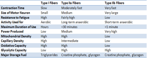
(Bushell, 2013)
The structure of a motor unit
A motor unit is made from a motor neuron and the skeletal muscle fibres innervated by that motor neuron’s axonal terminals (Purves, et al., 2001). A group of motor units is called a motor pool and the number of fibres in each unit can differ within muscles. This impacts precision and force generation. Differential initiation of single or multiple motor units with a motor pool can therefore control precision and force of movement.
Henneman’s size principle of motor unit recruitment
Henneman’s size principle states that; motor units are recruited from smallest to largest and as more force is needed, motor units are recruited in a certain order per the extent of their force output. This means that the smaller units are recruited first which means that it reduces the amount of fatigue an organism experiences by only using fatigue resistant muscle fibres, unless a higher force is needed and then fatigable fibres are used. This means that slow twitch, low-force, and fatigue resistance muscle fibres are activated before fast twitch, high-force, less fatigue resistant muscle fibres (Bawa, Jones, & Stein, 2014).
The motor unit and the Henneman’s size principle of motor unit recruitment
The structure of the motor unit
A motor unit is constructed from a motor neuron and skeletal muscle fibres, they innervated by the axonal terminals (Purves, et al., 2001). The motor neuron and its muscle unit are inseparable in function, this is because the action potetial in the neurons activates the fibres of the muscle unit (Karpati, 2010). A group of motor unit are gathered in columnar, spinal nuclei and this is called motor neuron pools. The number of fibres in each unit can differ from another and this then affects the force generation and the precision of the movement (Present, 1997).
The Henneman’s size principle of recruiting motor unit
The Henneman’s size principle expresses that motor units that are recruited from the smallest to the largest, this is because if more force is needed, then are recruited in a certain order due to the extent of their force output. Therefore, this means that the smallest motor units are employed first and this reduces the amount of fatigue that an organism experiences, by only using fatigue resistant muscle fibres, unless a higher force is needed, then fatigable fibres are used (Bawa, Jones, & Stein, 2014).
References
Al-Qusairi, L., & Laporte, J. (2011). T-tubule biogenesis and triad formation in skeletal muscle and implication in human diseases. Skeletal Muscle, 1(1). doi:10.1186/2044-5040-1-26
Bawa, P., Jones, K., & Stein, R. (2014). Assessment of size ordered recruitment. 8. Retrieved from https://www.ncbi.nlm.nih.gov/pmc/articles/PMC4112781/
Bushell, D. (2013). Muscle-specific hypertrophy: Chest, Triceps and shoulders. Retrieved from TheGymLifestyle: http://blog.thegymlifestyle.com/muscle-specific-hypertrophy-chest-triceps-shoulders/
Hwang, P. (2015). Targeting the sarcomere to correct muscle function. Nature Reviews Drug Discovery, 14(5). doi:10.1038/nrd4554
Page, M. (2001). Human body: An illustrated guide to every part of the human body and how it works. (A. Baggaley, Ed.) London: Dorling Kindersley Publishers.
Purves, D., Augustine, G., Fitzpatrick, D., Katz, L., LaMantia, A.-S., McNamara, J., & Williams, M. (2001). The Motor Unit. Sinauer Associates. Retrieved from https://www.ncbi.nlm.nih.gov/books/NBK10874/
Types of skeletal muscle Fibres. (2016). Retrieved from Ivy Roses: http://www.ivyroses.com/HumanBody/Muscles/types-of-skeletal-muscle-fibers.php
Cite This Work
To export a reference to this article please select a referencing stye below:
Related Services
View all


DMCA / Removal Request
If you are the original writer of this essay and no longer wish to have your work published on UKEssays.com then please click the following link to email our support team:
Request essay removal