Functions and Components of the Digestive System
| ✅ Paper Type: Free Essay | ✅ Subject: Physiology |
| ✅ Wordcount: 4458 words | ✅ Published: 23 Sep 2019 |
Table of Contents
Explain the function of each part of the digestive system identified in the diagram above.
Draw a diagram of a typical cell and state the function of each of its organelles.
Explain the difference between benign and malignant tumours.
Explain the structure and function of each element of the urinary system
Draw the structure of a nephron and explain how it produces urine
Name and explain three disease/disorders which affect the urinary system.
Introduction
In this assignment I will identify and describe the function of the main components of the digestive system. I will give a brief description of the digestive system and a diagram of the description will be inserted. I will identify the cell and its organelles along with giving the function of each organelle, a diagram will also be included. I will also identify the main tissues in the body, their functions and locations and an example of each tissue. Lastly I will identify and describe the main elements of the urinary system and three conditions that are related to the urinary system.
Part A
Explain the function of each part of the digestive system identified in the diagram above.
Digestion is the breakdown of food and other substances in the body. This breakdown happens when a group of organs such as; the mouth, oesophagus, stomach, small intestine, large intestine, appendix, rectum and anus join together to digest the food and other substances. The digestive system also consists of the accessory organs; gallbladder, liver and pancreas. This breakdown takes in all nutrients from food and then excretes the waste. There are four functions of the digestion system, for example;
- Ingestion: This is the take in of food.
- Digestion: This is broken down into two. Mechanical digestion and Chemical digestion.
Mechanical: This is done by breaking down the food physically such as mastication or chewing.
Chemical: Food is broken down by enzymes in the mouth called amylase.
- Absorption: This is when all food is processed and is passed through the body, all nutrients are absorbed through the blood and lymph capillaries and are then passed through the body.
- Elimination: All excess substances which could not be digested are excreted here through the bowel and therefore this forms faeces.
Mouth:
The digestion first begins here. When you take your first bite the chewing breaks the food down into smaller particles that can then be easily digested. The mouth then secretes saliva and begins mixing the food in your body which the body can easily intake.
Oesophagus:
The oesophagus is located near your trachea (windpipe). This is where food is received and travels down to your stomach by peristalsis. The oesophagus releases mucus to lubricate the passageway for food.
Stomach:
The stomach is a hollow organ and is C – shaped and here the first stage of digestion occurs here. The stomach has the ability to expand when it becomes full, a full stomach can hold up to 1 gallon of food. The cells in the stomach release and acid which processes and breaks down food even further, this is then released into the small intestine.
Small intestine:
The small intestine is 6 metres long and here is where 90% of digestion occurs. The small intestine has three parts: Duodenum, Jejunum and the Ileum. The walls of the small intestine contain villi and microvilli; these absorb the food and pass it through to the capillaries. The muscles of the small intestine contract and relax to pass food through to the large intestine, for further digestion.
Large intestine:
The large intestine is approximately 5 feet long and is arched shaped. It takes excess waste from the small intestine. The large intestine contains the rectum, colon, caecum and anus. The colon breaks down an remaining food with a bacteria that it contains.
Appendix:
There is no main purpose of the appendix but there have been studies to say that it dries out indigestible food by absorbing water and passes it through the capillaries and into the blood stream.
Liver:
The liver is the largest gland in the body, it is red-brown in colour, soft and wedged shaped. It is a vital organ as it produces bile, breaks down toxic substances such as alcohol and drugs, filters blood from the digestive systems and stores many vitamins (A, B12, D, E & K) and also stores glycogen (stores energy) and iron.
Gall Bladder:
The gall bladder is shaped like a pear and it is a sac which stores bile that is processed by the liver. Bile is a thick liquid as a result of the breakdown of RBC’s (Red Blood Cells).
Pancreas:
Shaped like a leaf or a fish, it is greyish/pink in colour and this has a very important role in our digestive system and the endocrine system. The pancreas is located behind the stomach. It is both an endocrine (an organ that produces hormones) and an exocrine ( an organ that produces enzymes) organ. It produces insulin and glucagon which is a hormone that is produced to regulate blood sugar levels and the amount of sugar we intake. It produces pancreatic juices which contain enzymes such as lipase (fat digestion), amylase (starch digestion) and trypsin (protein digestion).
Rectum:
This begins at the end of the large intestine and passes through to the end of the anus. The colon passes stool when it becomes full and pushes it through the rectum which then causes the bowels to move.
Anus:
The anus is an opening at the end of the body and this is where facies passes through and leaves the body.
Outline the composition of Proteins, Fats and Carbohydrates and explain how each are digested and absorbed by the body.
|
|
Proteins |
Fats |
Carbohydrates |
|
Composition |
They are made up of molecules of hydrogen, carbon, oxygen and nitrogen. They are composed of long chains of amino acids. Amino acids are the building blocks of proteins. |
Triglycerides are the dominant component of most fats. They are also composed of monoglycerides, + diglycerides, three fatty acids: phosphatides, starch and fat soluble vitamins. A triglyceride is composed of glycerol + 3 fatty acids. |
Composed of molecules such as carbon, hydrogen and o2. Molecules combine to form saccharides (sugar), the basic unit of carbohydrates. Single unit carbohydrates are called monosaccharides, Two units are called disaccharides and three or more units are called polysaccharides. |
|
How are they digested |
Pepsinogen is secreted in the stomach and is changed into pepsin due to the low PH condition. This then breaks down proteins so it can be absorbed by the body. An enzyme called trypsin is produced in the small intestine and this is where proteins are then further broken down with the use of this enzyme. This is then passed through the intestinal walls and sent into the capillaries. |
When fat is present in the small intestine it then produces hormones which stimulate the release of lipase which breaks down fats into monoglycerides and fatty acids. Bile breaks down fatty acids so that they can be absorbed. Short chains of fatty acids are absorbed directly into the blood through the intestine capillaries. Long chains of fatty acids are too large to fit into these capillaries so they are then absorbed into the fatty walls of the intestine. |
Amylase is present in the mouth where digestion begins but it is also present in the small intestine where it works with other enzymes to breakdown carbohydrates into monosaccharides. |
|
How are they absorbed |
These are absorbed by cells and used to build other proteins. |
Fat is absorbed in the small intestine and is then turned into triglyceride. |
Carbohydrates contain starch which is absorbed through the blood stream. |
Part B
Draw a diagram of a typical cell and state the function of each of its organelles.
Cell membrane: This is the outer layer of the cell; it is semi-permeable which means it only allows certain materials in such as O2, nutrients and water in and Co2 and waste products out. It protects the cell and gives it its shape.
Nucleus: This is the brain of the cell and it contains chromosomes and DNA, this part of the cell plays a part in single cell division (mitosis).
Nuclear membrane: This separates the nucleus from the cytoplasm and protects the nucleus.
Cytoplasm: A jelly like substance, which holds all organelles in place.
Mitochondria: The power house to the cell, this provides energy for the cell. It breaks down food and releases energy.
Golgi Bodies: These are the communication centre for the cell. It stores energy and secretes it around the cell.
Ribosomes: These make protein for the cell.
Lysosomes: These digest waste in the cell.
Classify tissues into four main groups; epithelial, connective, muscle and nervous, give an example of each?
Epithelial Tissue
This type of tissue is widespread throughout the body, it helps protect, absorb and secrete products in the body. It is a type of tissue that is made up of closely packed cells that are arranged in continues flat sheets. They have on free surface and do not contact any other cells. It has no blood supply but has an excellent nerve supply. The cells in this tissue have the ability to renew themselves.
|
|
Function |
Location |
|
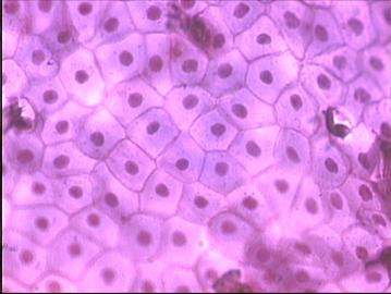
Simple squamous:
|
This is a single layer of flat cells in contact with the basal layer. This type of epithelial tissue is permeable and this occurs when small molecules need to pass through membranes through filtration. This tissue is very thin and provides little protection |
This type of tissue is located in the capillaries, alveoli, glomeruli and the outer layer of the skin. |
|
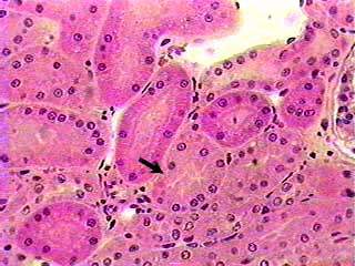
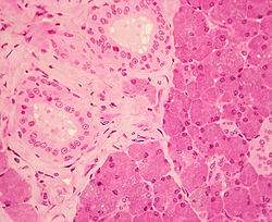
Simple cuboidal:
|
Consists of single layered, cube cells. These cells provide protection and they may be active or passive depending on the location. |
Usually found in the kidney tubules, liver, thyroid, mammary and salivary glands. |
|
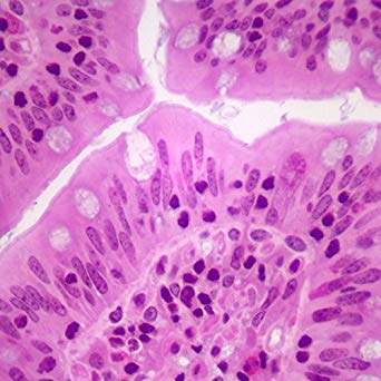
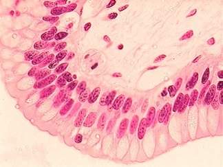
Simple columnar:
|
This type of tissue is made up of a single layer of epithelial cells, this often occurs when we see regions where secretion and absorption happens. These cells are arranged in a neat row where then they form to create the simple columnar tissue. Its main function is for protection. |
Found in the oesophagus, stomach, small intestine and the large intestine. |
|
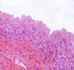
Transitional:
|
This tissue has cells stretch in order to accommodate the levels of fluid in an organ. |
These are found in the bladder and ureters. |
|
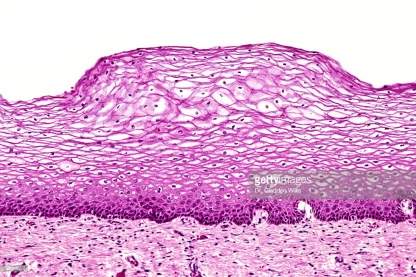
Stratified Squamos epithelium:
|
The cells here are flat on the surface and contain no source of activity. These types of cells have no nucleus or no organelles. They are mainly filled with keratin which nourishes the skin. |
These are found in the epidermis layer of the skin, the palms of the hands and soles of the feet. |
|
Stratified columnar epithelium:
|
These cells usually consist of several layers; the basal cells are cuboidal in shape and the superficial cells are elongated and columnar. Their main function is to protect the skin and secretion of waste products out of the skin. |
Small amounts of these cells can be found in the urethra and in the ducts of some glands. |
|
Stratified cuboidal epithelium:
|
These are made up of cube shaped cells that are used as protection to the skin. |
Found in the bladder and urinary tract. |
Muscle Tissue
This type of tissue contracts and relaxes and there is three main types:
Skeletal
Smooth
Cardiac
|
|
Function |
Location |
|
Cardiac |
Cardiac muscle tissue allows blood to be pumped to all areas and organs of the body. It used by involuntary movement and it never rests. |
Only found in the heart. |
|
Smooth: |
Smooth muscles tissue forms body organs and can change its shape to suit its functions. Under involuntary movement and peristalsis occurs here. |
Found in the hollow organs e.g. stomach and bladder. |
|
Skeletal: |
Under voluntary movement and here this type of muscle tissue allows body movement of the bones. |
Found attached to bones. |
Nerve Tissue:
Nerve tissue consists of neurons and neuroglia these are in the brain, spinal cord and nerves. These neurons send impulses to the brain and spinal cord they need a constant supply of oxygen and glucose.
|
Function |
Location |
|
|
Neurons: |
These send impulses from the brain and to the body and then to the spinal cord. Neurons have 4 different types of impulses that are connected to it. Dendrites – These receive the information. Axon – This sends the information to the neurons Myelin – This is the protection layer of the axons, it also insulates them. Synapse – This transmits the messages from one neuron to the next. |
Found in the brain and spinal cord. |
|
Neuroglia: |
The neuroglia protects and provides nourishment to the neurons. |
Found in the tissues of the brain and spinal cord. |
Connective Tissue:
Connective tissue connects all tissues in the body. Connective tissue supports and protects the body. It varies in blood supply especially in ligaments which is why it takes a lot longer for them to heal. There is three main types of connective tissue:
Solid
Semi-Solid
Liquid
|
|
Function |
Location |
|
Cartilage: |
Cartilage is a form of semi-solid tissue. Cartilage forms the outer ear, moves joints in the body and moves the vocal cords. |
Found in the ear, larynx and trachea. |
|
Bone: |
The bone in the connective tissue is made up of collagen fibres. Bones can either be dense or cancellous. Bones contain osteoblasts and osteocytes cells |
Ligaments. |
|
Adipose: |
Adipose is another term for fat. This type of connective tissue is used as a protective layer due to its fatty, cushion like ability. Adipose connective tissue is made up of closely packed cells. |
Can be found in bone marrow and surrounding the internal organs. |
|
Blood: |
Blood is a type of liquid connective tissue, it contains three types of cells; erythrocytes, leukocytes and thrombocytes. The main function of blood is to transfer nutrients, oxygen (O2), hormones and removes waste. |
Circulates the cardiovascular system. |
|
Lymphatic: |
This is another type of liquid connective tissue. It is responsible for the manufacturing of blood cells in the body. It is formed of leukocytes and soluble liquid proteins that are made during clotting. |
Lymphatic system |
|
Elastic |
This type of tissue is made up of elastic fibres from chondrocytes cells. It helps maintain homeostasis through maintaining blood pressure and allows normal exhalation. |
Found in the skin and lungs. |
|
Fibrous: |
This type of connective tissue is made up of fibroblast cells and is made up of fibrous fibres. It gives strength to the inner layer of skin and gives movement to joints. |
Found in the dermis layer of the skin and in ligaments. |
(htt)
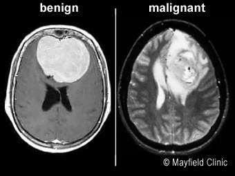
Explain the difference between benign and malignant tumours.
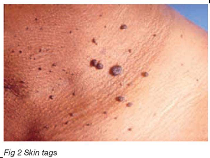
Tumours are a group of cells that join together and multiply or divide in an abnormal way, forming a lump or mass in or on the tissue. There are two type of tumours; Benign and Malignant tumours.
|
Benign |
Malignant |
|
Non-Cancerous |
Cancerous |
|
Do not spread or invade to other organs |
Can spread or invade to other organs and tissues |
|
Can cause pressure or pain and can push organs out of place |
They travel through the lymph and blood this is the process of metastases |
|
Enclosed in a capsule |
Damages the tissues and organs surrounding the tumour |
|
Can be surgically removed |
Increases in size |
|
Usually do not re-occur |
Often can re-occur |
|
Example of a benign tumour would be warts, moles or skin tags.
|
Example of malignant tumour would be ovarian cancer, prostate cancer, pancreatic cancer, mouth cancer. |
Part C
Explain the structure and function of each element of the urinary system
The urinary system is a system which consists of many organs in the lower part of our bodies, such as the kidneys, ureters, the bladder and the urethra. The urinary system has many important functions which help us secretes/ removes waste and excess fluid from our body, filters our blood, regulates our fluid levels. The urinary system also produces a hormone that also controls the rate at which our blood cells create, this hormone is called Renin. It produces an enzyme that controls our blood pressure. This helps the body to remain in balance or otherwise called homeostasis. Homeostasis is the maintaining of the bodies internal balance.
Kidneys:
The kidneys are bean shaped and are red in colour. They are located just towards the bottom of your back where your hips are located. They are enclosed in a clear membrane called the renal capsule and this is used as protection of infection for the kidneys. The kidneys allow blood to be passed through for it to be cleaned every day the renal artery delivers this blood to the kidneys and it is here where 180litres of blood is passed through. After this process is done the blood is filtered and sent back to the heart by the renal vein.
Ureters:
These are two slender, muscular tubes that pass the urine from the kidneys to the bladder. They are wider at the top and are narrow at the bottom. The ureters are lined with a mucous membrane, and this aids to fight infection. The walls of the ureters contract moving the urine away from the kidneys and towards the bladder.
Bladder:
This is a hollow sac that holds the urine. The bladder is lined with a mucous membrane and contains three layers of smooth muscles tissue on the walls of the bladder. The internal sphincter keeps the bladder closed while it begins to fill. The external sphincter allows the muscles to relax and this is when urine is passed through, the bladder can hold up to 1 pint of urine.
Urethra:
The urethra is a tube that carries urine from the bladder to the outside of the body. Male and female urethras have different functions and compare in size. Females urethras only main purpose is to pass urine and is approximately 1 ½ to 2 ½ inches long. Males urethra is used to pass urine and semen and is approximately 6-8 inches and is S – shaped.
Draw the structure of a nephron and explain how it produces urine
The nephron is a structure in the urinary system that produces urine. It is a long tubule which is approximately 1.2 inches long. It consists of the renal corpuscle, capillaries (glomerulus), the Bowman’s capsule, tubules, collecting duct and loop of Henle. It goes through a process to produce urine for example:
Filtration
Reabsorption
Secretion
(htt1)
- There are 1 million tiny nephrons in each kidney which clean the blood that flows through the kidneys.
- Each tiny nephron contains glomerulus and tubules. The glomeruli have capillary beds which are found in the Bowman’s capsule.
- These capillaries have walls that have tiny openings in them, due to the blood being a lot of pressure, the liquid of the blood pushes through the walls of the capillaries, which then removes the waste and the nutrients are in it.
- The liquid then goes into the tubules and these then make up the nephron. This liquid is called the filtrate. This is the process of filtration.
- The capillaries absorb the substances that it can make use of such as nutrients, water and mineral back into the blood stream. This process is known as reabsorption.
- When the filtrate reaches the end of the tubules, the only product left is excess fluid and waste which is known as urine. The urine is emptied into the renal pelvis (collecting area for urine) and from here the urine flows into the ureters. This is known as secretion.
Name and explain three disease/disorders which affect the urinary system.
Urinary Tract Infection (UTI):
These are one of the most common types of disorders or infections that can occur in an part of the urinary system. They are most common in woman than they are in men. UTI’s can be very painful to the person who may be infected. The infection begins when tiny microorganisms attach themselves to the opening of the urethra and the tiny microorganisms begin to grow. Urine is usually clean and free from bacteria but when these microorganisms occur this can cause a urinary tract infection. A urinary tract infection can become dangerous if it spreads to the kidneys and this can then lead to a kidney infection or even more life threatening, sepsis.
Sometimes urinary tract infections do not show any signs or symptoms but when they do they generally show up as;
Burning when urinating.
When there is a sign of blood in the urine.
A pungent smell in urine, as urine should have no smell.
Pelvic pain.
Urine seems to become cloudy.
Discharge.
UTI’s are usually treated with an anti-biotic but if not treated in the correct procedure it can cause complications to the body’s urinary system. For example; recurrent infections, chronic kidney infections if not treated at all, sepsis if the bacteria spreads to the kidneys.
Some people can be more prone to getting UTI’s and they are completely normal, as they are the most common infection. Anything that can block the flow of urine leaving the body can cause a UTI or anything that can slow the flow of urine can lead to a risk of infection.
Kidney Stones:
Kidney stones are when the salts in the urine can form solid crystals. Kidneys stones obstruct the flow of urine out of body can then cause infection, kidney damage or kidney failure. Kidney stones do not cause much damage to the urinary system if they are caught in early stages.
They can cause a lot of pain usually medication is given for this pain and people are advised to drink more water. If the kidney stones become trapped in the urinary tract, surgical removal of the stones may be required.
There are different types of kidney stones for example:
1) Calcium stones: The most common type of kidney stones. These are formed from calcium that is not being used by the muscles and bones, and it then forms into a build-up which then forms the calcium stones. These are combined with oxalate and phosphate. This type of kidney stone can occur from in taking certain medications like medication for seizures or migraine medication.
2) Struvite stones: These are caused when there is an infection in the urinary tract. Usually UTI’s can be the main cause of these stones.
3) Uric acid stones: These happen when there is too much acidity in the urine. People who do not drink enough fluids can usually get these.
4) Cystine stones: These are more a rare type of kidney stones but can be hereditary.
Bladder Incontinence:
Incontinence is when a person loses control of their bladder and the nerves in the urinary system do not send impulses to the brain letting them know when their bladder is full, this then leads to a person urinating themselves. It is a common condition but usually occurs in elderly people.
There are different types of incontinence such as:
- Stress incontinence: This happens when a person accidentally releases urine when there is stress being put on the bladder. Such stress can occur as sneezing, coughing laughing or exercising.
- Physical incontinence: This can happen when a person with a physical condition does not make it in time to the toilet and then loses complete control of their bladder. Somebody who may have arthritis might find it difficult to open the button of their trousers in time.
- Overflow incontinence: This happens when a person’s bladder does not empty in the correct way, so it releases dribbles of urine.
Some medications, foods, drinks or alcoholic drinks may have a cause to temporary incontinence; these stimulate your bladder making your bladder full, these can include:
- Alcohol
- Large doses of vitamin C
- Chocolate
- Caffeine
- Muscle relaxants or sedatives
Other causes for bladder incontinence can be from UTI’s which are more common in women, if these are not treated in the correct procedure these can cause bladder incontinence. Prostate gland enlargement can cause loss of control of the bladder in men, pregnancy and childbirth is another factor that can cause loss of bladder control, menopause, and change of age.
(htt4)
Conclusion
In this assignment I described the main functions of the digestive system. I gave a brief description of what the digestive system is and gave the structure and function of the main components of the digestive system. I named the different types of organelles in a cell and gave each of their functions. I further went on to explain the different types of tissues, their function and location and also a brief description of the function of each tissue. I ended my assignment off with the urinary system. I outlined the main elements of the urinary system and each of their functions along with three conditions related to the urinary system.
Works Cited
- (n.d.). Retrieved from https://sciencing.com/7-types-connective-tissue-8768445.html
- (n.d.). Retrieved from https://www.britannica.com/science/nephron
- (n.d.). Retrieved from https://www.mayoclinic.org/diseases-conditions/urinary-tract-infection/symptoms-causes/syc-20353447
- (n.d.). Retrieved from https://www.mayoclinic.org/diseases-conditions/kidney-stones/symptoms-causes/syc-20355755
- (n.d.). Retrieved from https://www.mayoclinic.org/diseases-conditions/urinary-incontinence/symptoms-causes/syc-20352808
- Class Notes
Cite This Work
To export a reference to this article please select a referencing stye below:
Related Services
View all


DMCA / Removal Request
If you are the original writer of this essay and no longer wish to have your work published on UKEssays.com then please click the following link to email our support team:
Request essay removal