TIRF Microscopy for Counting Molecules in Bacterial Flagellar Motor
| ✅ Paper Type: Free Essay | ✅ Subject: Physics |
| ✅ Wordcount: 1388 words | ✅ Published: 09 Mar 2018 |
How TIRF microscopy has enhanced the way single molecules are counted in the bacterial flagellar motor
Abstract
The counting of individual molecules is important in order to establish how many molecules there are in a particular system. TIRF microscopy is one method to count molecules. The bacterial flagellar motor is a complex system in which motility protein B molecules can be counted using TIRF. Discussion is given for a key research topic based on counting of molecular subunits of this motor. Reviews of the background areas, limitations and confirmations of this research are conducted, and a discussion of the research and its contributions to technological and medical applications.
Introduction: The bacterial flagellar motor, TIRF microscopy and associated research
Flagellar motors are machines used to drive many bacteria which have to swim in a solution like our bodies. This motor, usually studied in E. coli bacteria, is powered by a flux of H+ or Na+ ions across a cytoplasmic membrane driven by an electrochemical gradient (Sowa and Berry, 2008). The motor itself consists of two components, a rotor and a stator: the rotor spins relative to the cell and is attached by a helical filament known as a hook, whereas the stator is fixed to the cell wall (Francis et al 1994).
A method commonly used to visualise the bacterial flagellar motor is Total Internal Reflection Fluorescence (TIRF) microscopy, which is one of the most frequently employed methods in bio-optical research (Leake 2013, P87). TIRF microscopy uses an evanescent field to illuminate the area covered by the specimen in question, which is adjacent to a glass-water interface. Using organic dyes has made it possible to view other properties of bacteria using TIRF (Sako et al 2000). This method is useful in counting the molecular subunits of the bacterial flagellar motor.
TIRF microscopy has been used to view single molecules within live bacteria. For viewing the bacterial flagellar motor of E. coli, scientists tagged motility protein B (MotB) cells with Green Fluorescence Protein (GFP) in order to detect them via TIRF. This highlighted the areas within the bacteria where the motor was situated. To visualise the bacteria in a single confined position, the cell was tethered to the slide for viewing on the microscope. This is shown in Fig. 1, where the fixed position of the flagellum limits the bacteria’s movement to rotation.

Fig. 1 Tethered cell showing its exposure to the evanescent field used for TIRF (Leake 2006, P355)
Background, difficulties and discoveries from the research
The history behind counting molecules
Though the basis of this experiment began in the 60s, initially using the measurement of the activity of single molecules (Rotman 1961), optical detection and spectroscopic methods are now used instead. The counting of complex molecules can now also be achieved, but this area of research also needs TIRF microscopy. TIRF was enhanced in 1984 by Daniel Axelrod after the publishing of a paper on its experimental methods (Axelrod 1984), and those methods remain largely unchanged today. Furthermore, GFP molecules have only been recently understood. Without this research and development in GFP, visuals using TIRF would not be possible (Tsien 1998).
The difficulties encountered and overcome in counting molecules
An estimate of around twenty-two molecules are thought to be present in the flagellar motor, with roughly eleven stator units. The main issue with determining this result explicitly is that there are many MotB molecules not associated with the motor. These molecules cause a problem as they are free to diffuse within the motors of the cell membrane. The fluorescence intensity was estimated from the areas where it was clear that no such molecules would interfere with results. Additionally, an intense laser beam focus for TIRF was required to photobleach GFP molecules. Only an extremely small region of the bacteria was viewed to improve the ability to track a small number of molecules – a significant amount of noise remained in the system, however, meaning that it is not yet possible to count exactly how many molecules are in each motor.

Fig. 2 showing bright field (top) and their corresponding TIRF images (bottom) (Leake 2006, P355). The bright areas represent the flagellar motor.
Using TIRF, bright spots indicate the centre of the cell rotation of the image shown in Fig. 2. There was a high density of spots centred on the flagellar motor, due to the high density of GFP-MotB molecules around the motor. Short times (between 0-10 seconds) are used because TIRF illumination over the bright spots decreases over longer periods of time, which makes it difficult to detect regions of the flagellar motor. Care was taken to not cause damage to the GFP due to the excitation light on the surrounding water: this means that smaller time steps were required such that the GFP molecules emitted a constant amount of photons.
The effects FRAP and FLIP
With the noise effects reduced, there was the opportunity for the GFP-MotB molecules to spread into the area which had been bleached beforehand. Focusing the laser beam onto the motor itself resulted in the effects of fluorescence recovery after photobleaching (FRAP) and fluorescence lost in photobleaching (FLIP). Observations of the molecular turnover in the cell found that over a period of five minutes, the intensity of the bright spots around the motor would decrease to nearly zero but then recover to half the initial intensity. This implied that binding and unbinding at the motor and bleaching occurred in the evanescent field (Leake et al 2006, P357), which means that the stator units in the motor only spend half a minute in each flagellar motor. This is demonstrated in Fig. 3, which illustrates the time elapsed after laser focused bleaching and how the intensity decreases but eventually recovers.
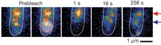
Fig. 3. Shows the effects of FLIP and FRAP over the period of 5 minutes (Leake et al 2006, P357)
Benefits of the research and potential uses for the future
An improved type of MotB was used in the research, which enhanced the way in which the molecules in the motor were counted. FLIP and FRAP indicate an alternative means for visualising the motor in motion, confirming that the stator units are dynamic instead of static (Sowa and Berry, 2008, P117). This is one of the first measurements of turnover in a molecular machine, establishing other possible characteristics which could be exploited to gain further understanding of the motor (Leake et al 2006, P357).
Scientists are keen to understand more about how such motors work, so that developments in the delivery of medicine or for environmental purposes can be made. It may be possible to replicate the motor (Fukuda et al 2012). Delivery of medicine is one of the key goals: modelling the bacterial flagellar motor such that it could be used for targeted drug delivery would be revolutionary (Leake 2013, P259). Furthermore, through the development of nano-bots, this could be used to visualise diseased tissue or uncover parts of the human body.
Summary
This area of biophysics is relatively new: from the discoveries in the early 60s through to the 80s, there has not been a clear link between the two subjects. From the late 90s there was an opportunity to visualise biological material using physical optical devices. Over the past two decades, it has now reached to the point where it is possible to count single molecules to a close estimate. The use of GFP molecules combined with TIRF can enhance the visualisation of molecules in bacteria, and there are methods which can significantly improve the estimation of the number of molecules in the motor. This is still a difficult process due to the interference of other, unrelated molecules. FLIP and FRAP methods have proved that the stator is a dynamic rather than a static component of the motor. There are specific parts of this research which may be useful for future technological applications, for example: the delivery of medicine or the bio-sensing of diseased tissue.
References
Axelrod, D; Ann. Rev. Biophys. Bioeng.; 13; 1984; 247-68
Francis, N, R.; Sosinsky, G,E; Thomas, D; Derosier, D. J; Journal of Molecular Biology 235, 1994; 1261–1270.
Fukuda, T; Kojima, M; Zhang, Z; Nakajima, M; Biomed Micro-device; 2012; 1027-32
Leake, M; Single Molecular Cellular Biophysics; 2013
Leake, M C; Chandler, J H; Wadhams, G H; Bai, F; Berry, R M; Armitage, J P; Nature 443; 2006; 355-358
Rotman, B; Biochemistry 47; 1961; 1981-91
Sako, Y; Minoghchi, S; Yanagida, T; Nature Cell Biol. 2; 2000; 1929-1932
Sowa, Y; Berry, R, M; Quarterly Reviews of Biophysics 41, 2008, 103-132
Tsien, R.Y; Annu. Rev. Biochem 67; 1998; 509–44
Cite This Work
To export a reference to this article please select a referencing stye below:
Related Services
View all


DMCA / Removal Request
If you are the original writer of this essay and no longer wish to have your work published on UKEssays.com then please click the following link to email our support team:
Request essay removal