Systems in the Human Body
| ✓ Paper Type: Free Assignment | ✓ Study Level: University / Undergraduate |
| ✓ Wordcount: 9602 words | ✓ Published: 06 Jun 2019 |
Audrey Supan
Main Organs
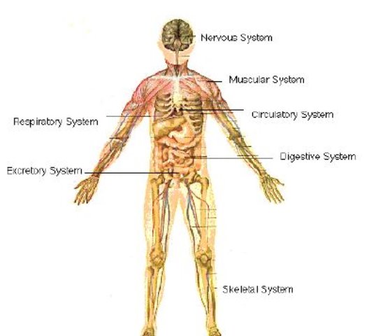
The human body systems work together which performs essentially to maintain health. Most parts of the human body rely on each other to make sure they all function properly to remain stable. Some organs may be part of more than one body system, if they serve more than one function. All body systems are important for life to be sustained.
There are 15 main organs in the body that helps in everything a human does on a day-to-day basis. These are the skin, heart, lungs, stomach, bladder, brain, eyes, ears, pancreas, intestines, liver, ovaries, kidneys, testes and uterus.
The Skin
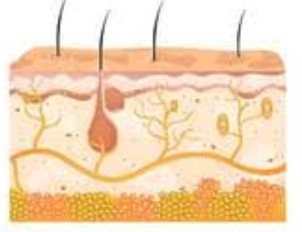
The skin is the outer layer that protects everything else internally and it is made up of different types of tissues. Its role is to help external stimulation, fights against damage or infection, prevents drying out, helps with the regulation of body temperature, excretes waste such as sweat, stores fat and makes Vitamin D*. It holds many tiny structures, each with different function. The whole skin which includes tissue layers and structures is called the integumentary system.
The three types of layers of the skin are the epidermis, dermis and the subcutaneous layer. The epidermis is the thinner layer of the skin which forms epithelium (a thin tissue that forms the outer layer of the body’s surface and lining the alimentary canal and other hollow structures). Whereas, the dermis is the thick layer of the connective tissue under the epidermis and it contains many capillaries that supplies food and oxygen. And lastly, the subcutaneous layer is a layer of fatty tissues which is found below the dermis which stores larger blood vessels and nerves. It acts as an insulator and maintains a balanced body temperature.
There are 8 structures in the skin and these are Meissner’s corpuscles, sebaceous glands, Arrector Pili Muscle (hair erector muscles), hair follicles, pain receptors, hair plexuses, sweat glands and Pacinian corpuscles.
Meissner’s corpuscles are a type of nerve ending in the skin which is mostly responsible for sensitivity to light touch. This can be found mainly on the fingertips and palms. Its role is to send impulses to the brain when the skin comes in contact with an object.
Sebaceous glands are tiny glands in the skin which opens into hair follicles and produces oil called sebum which waterproofs the hairs and epidermis, keeping them supple. As dead outer cells are difficult for microbes to penetrate, the oils from the sebum help kill microbes.
Arrector Pili muscle is a small muscle that connects to each of every hair follicles and the skin of the body. When you get cold, your blood vessels constrict which straightens the hairs so that it can trap warm air and improve insulation; this also cause goosebumps. However, when we get too hot, our blood vessels dilate so that it allows more warm blood to flow near the surface of the skin, where the heat can be lost to the air and this causes us to sweat which also help cool us down.
The hair follicles are long, narrow tubes and all contains hairs. This grows new cells which add to its base from the cell lining the follicle. The older cells die as keratin and are formed inside the hairs.
Pain receptors are found in nerve fibre endings in the tissue of most inner organs and also in the skin. They are receptors that send impulses to the brain when any stimulation becomes excessive such as pressure, heat or touch. This is what causes a sensation of pain.
Hair plexuses is a special group of nerve fibre endings and each form a connection around the hair follicle which is a receptor that sends nervous impulses to the brain, in this case when our hair moves.
Sweat glands excrete sweat and each has a narrow tube called the sweat duct going to the surface. Sweat consists of water, salts and urea which enter the gland from the cells and capillaries.
Pacinian corpuscles are formed around single nerve fibre endings which lie in the lower skin layers and in the walls of inner organs. It is a pressure receptor that sends impulses to the brain when tissues receive deep pressure than light touch.
The Heart
The heart is a muscular organ which pumps blood around the blood vessels other known as the cardiovascular system. The heart is mostly hollow and it is composed of cardiac muscles called myogenic and connective tissues.
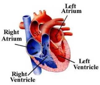
There are four valves in the heart which stops blood from going the wrong way and they are placed between the right atrium and ventricle, between the left atrium and ventricle, at the entrance to the pulmonary artery and at the entrance to the aorta.
The atria are the two upper chambers; the left side receives oxygenated blood from our lungs through the pulmonary veins. Whereas, the right atrium receives the deoxygenated blood from the rest of the body.
The ventricles are the lower chambers; the left ventricle receives blood from the left atrium which pumps into the aorta. Whereas, the right ventricle receives blood from the right atrium which pumps through the pulmonary trunk to the lungs. The ventricles contracts which forces blood to come out.
The aorta is the largest artery in the body; its purpose is to carry blood with fresh oxygen out of the left ventricle to start the process round the body. The pulmonary trunk is an artery that carries blood needing fresh oxygen out of the right ventricle. When it has left the heart, it separates both ways in the pulmonary arteries, one going to each lung. Whereas, the four Pulmonary veins carry fresh oxygen from the blood to the left atrium. The right pulmonary veins come from the right lung whereas, the two pulmonary veins come from the left lung.
The muscular wall of the left ventricle is thicker than the right side as it has to pump blood around the body. The right ventricle is thinner as it only needs to pump blood through the lungs. The heart has its own blood supply which comes from the coronary arteries.
Heart is the most vital organ which locates in the centre of the chest with a slight tilt to the left. With rhythmic muscle contractions, it helps circulate blood through the veins and arteries that supplies tissues with oxygenated blood and get rid of waste.
The cardiac cycle is the process of making up one complete pumping action of the heart other known as heartbeats which is about 70x a minute. The first process is when the two atria contracts and pumps blood into the ventricles which relax in order for it to receive it. When the atria have relaxed, it takes in the blood, which the ventricles pump out by contraction. The different valves open and closes during the cycle just so blood loss won’t occur.
Respiratory System
The respiratory system covers three processes in ventilation, external and internal respiration. Ventilation is all about taking in oxygen and expelling carbon dioxide, external respiration is the exchange of gases between the lungs and the blood. Lastly, the internal respiration is the breaking down of food, using oxygen and producing carbon dioxide.
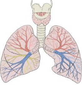
Both lungs are sponge like, air- filled organs which are located on either side of the thorax. Each lung is split into lobes, the right has three and the other only has two therefore allows more space to accommodate the heart. The tissue in the lungs is almost as 40 times bigger than the body’s outer surface which makes the lungs one of the largest organ in the body. The lungs are one of the most hard-working organs in the body. They
Expand and contract up to 20x a minute so they can supply oxygen to be distributed to tissues all over the body and expel carbon dioxide that has been created throughout the body.
Breathing is an automic which stems from the brain and it is situated in diencephalon. This is made up of inhalation and exhalation, both actions are automatic which is often controlled by nerves from the respiratory centre in the medulla which is found in the brain that controls many unconscious actions. This act when it detects too high a level of carbon dioxide in the blood.
Inhalation is the act of breathing in which causes the diaphragm contracts and flattens, lengthening the chest cavity. The muscles between the ribs also contracts, pulling the ribs up and outwards which help widen the cavity. This is so the expansion can lower the air pressure in the lungs, and then the air rushes to fill in the lungs.
Exhalation is the act of breathing out which causes the diaphragm and intercostal muscles to relax so that the air can be forced out of the lungs as the chest cavity decreases in size.
- Carbon dioxide exits


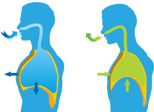
- Oxygen in


- Intercostal muscles relaxes, ribs move down and inwards
- Intercostal muscles contract, pulling ribs out and outwards
- Diaphragm relaxes
- Diaphragm flattens
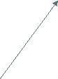
 Trachea is the main tube in which is responsible for passing air on its way to and from the lungs. The lungs are the two main breathing organs which gases are exchanged and they contain a lot of tubes and air sacs called the alveoli. Alveoli’s role is to allow oxygen to dissolve in its moist surface and this has a thin lining for an easier diffusion of gases. It also got a dense channel of blood capillaries for easy gas exchange and a large
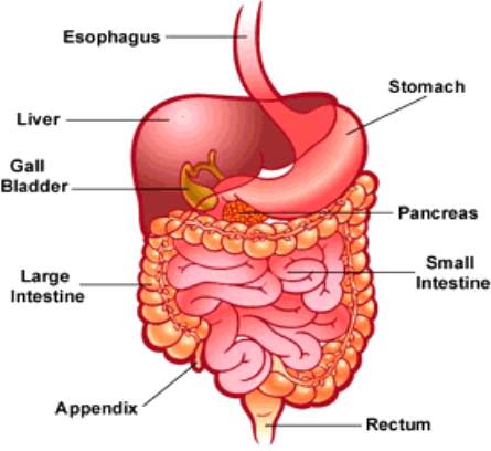
The Digestive System
After the food has been ingested, it goes through the digestive system; this then breaks down into soluble substances by a process called digestion. The substances will then get absorbed into the blood vessels around the system then transported to the body cells. The pancreas and liver play a vital role in digestion
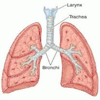
The stomach is a J-shaped, elastic, hollowed organ which is located just inferior to the diaphragm in the left part of the abdominal cavity. It is located between the oesophagus and the duodenum, the stomach is like a roughly crescent-shaped enlargement of the gastrointestinal tract.
The oesophagus or a gullet is the tube which food travels down to the stomach. The duodenum is the first part of the small intestine immediately beyond the stomach, leading to the jejunum. There are two types of sphincter which are cardiac and pyloric. Cardiac sphincter is a muscular ring between the oesphagus and stomach; this relaxes so that food can go through. The pyloric sphincter however is a muscular ring between the stomach and the small intestine; this relaxes once after a change have occurred so that food can go through.
The inner layer of the stomach is full of wrinkles known as rugae which allow the stomach to stretch in order to take in larger meals and help grip and move the food during digestion. The size of the stomach may come differently depending on the person, but on average it can comfortably contain 1-2 litres of food and liquid during a meal. When this stretch, it can hold up to 3-4 litres when we overeat or have larger meals.
Through the process of digestion, the stomach produces hydrochloric acid. This helps kill off many harmful microorganisms that might have been swallowed along with the food. This is important as enzymes in the stomach work better in acidic condition at a low ph. The stomach can store a meal for about 1-2 hours. During this period, the stomach continues the digestive process that started in the mouth and lets the intestines, pancreas, gallbladder, and liver to prepare to complete the digestive process.
The food goes through four processes once masticated. Firstly, it is ingested which is the process of putting all nutrients in your system. Secondly, it gets digested, which is the mechanical and chemical of breaking down of food then this goes through the process of absorption which is when all digested food gets absorbed making it to waste. Lastly, it gets eliminated which gets rid of all unneeded materials through excretion.
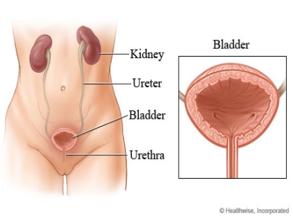
The Urinary System
This is the main system of body parts which involves in excretion; this is the expulsion of unneeded substances. The lungs and skin are also involved in excretion which expels carbon dioxide and sweat respectively. The bladder is a sac that holds up urine. Its lining has many folds which is called rugae, this flattens out as it fills up, making it bigger. This is located in the lower abdominal area near the pelvic bones
The bladder, like the stomach, is an expandable saclike organ that contracts when it is empty. The two muscular rings, the internal and external urinary sphincter controls the opening from the bladder into the urethra. When liquid reaches up to a certain level, nerves will start to stimulate the internal sphincter to open while the external sphincter stays under conscious control and be closed for longer period of time.
The urethra is the tube carrying urine from the bladder out of the body. This process is called urination. The urine is the liquid which exits the kidneys. Its main components are excess water, urea which is a nitrogen- containing waste material which help with process of breaking down excess amino acids in the liver. It travels through the blood to the kidneys, along with smaller amounts of similar substances.
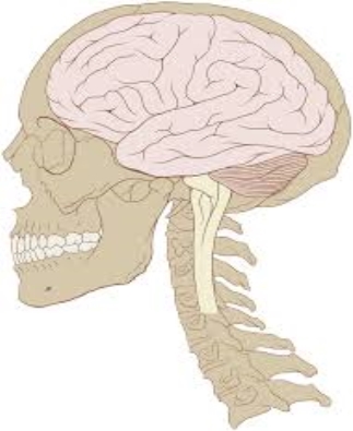
The Brain
This is an organ which controls most of the body’s activities. It is the only body organ that is able to produce intelligent action based on stored information, this includes present events and future plans. It is made up of vast amounts of neurons, arranged in sensory association and motor areas. The sensory areas receive the information from all body parts whereas the association areas analyses impulses and make decisions. The motor areas send impulses to the muscles or glands. These impulses are carried by the fibres of 43 pairs of nerves in the spinal cord.
The human brain is the largest brain of all vertebrate’s relative to body size, it weighs about 3.3 lbs. and it makes up about 2% of a human’s body weight. The cerebrum makes up 85% of the brain’s weight and contains about 86 billion nerve cells called the “grey matter”. It contains billions of nerve fibres which are axons and dendrites; “white matter”. These neurons are connected by trillions of connections, or synapses.
The Cerebrum, being the largest area is made up of two halves. The right half controls the left side of the body and the left side controls the right. Each half splits into four lobes which controls different functions like the thinking, memory, behavior, personality, learning, hearing, sight, language and touch. The cerebellum is the area which co-ordinates muscle movements and balance, the two things under the overall control of the cerebrum. This is located at the back of the brain. The brain stem controls vital functions such as breathing and heart rate and the pituitary gland is at the base of the brain which controls hormone production in the body.
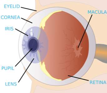
The Eyes
The eyes are the organs for living organisms to be able to see, this sends nervous impulses to the brain when stimulated by light rays from external objects. The brain interprets the impulses to produce images. Each eye consists of a hollow, spherical capsule which known as the eyeball. This is made up of different layers and structures. It is placed into a socket in the skull and it is protected by the eyelids and eyelashes.
The eyes have several major components which are the cornea, pupil, lens, iris, retina and sclera. These organs all work together to capture an image and transmit it directly to the brain’s occipital lobe through the optic nerve. Eyes are approximately one inch in diameter. Pads of fat and the surrounding bones of the skull protect them.
Our eyes can only see in two dimensions, we are able to determine distances and depth in our three-dimensional world. This is so the brain can interpret the two slightly different images our left and right eyes visualise as one. This is called stereoscopic vision. Other visual cues like shadows, how objects are blocking each other, and our knowledge about the sizes of different objects also help us determine depth and distance.
When a person looks at an object, the light that reflects from it enters the eyes and then refracted or bend so that it can create a focused, upside down image of the object which the brain could interpret and turn to a right direction. In the actual eye are photoreceptors which create nerve impulses when struck by light. These are two types: cones make colour vision possible, and rods specialise in black and white images.
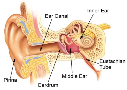
The Ears
The two ears are the organs of hearing and balance, but this depends on specialised receptors called hair cells. Each one is split into three parts, one being the outer ear, second being the middle ear and the third the inner ear.
The outer ear is the shell of the skin and cartilage, together with a short tube and this tube lining has its own special sebaceous glands which secrete ear wax. The middle ear is an air- filled cavity that contains a chain of three tiny bones called the malleus and stape. Lastly, the inner ear is a connected series of cavities in the skull, with tubes and sac inside them.
Through the process of hearing, the eardrum vibrates when soundwaves enter the ear canal. Ossicles are the three tiny bones that are found in the ear which also includes the stapes, being the smallest bone in the body. Vibrations pass to the oval window, which is a membrane at the entrance of the inner ear. Sound waves enter through the outer ear, and then it travels down to the middle ear till it reaches the inner ear and its intricate network of nerves, bones, canals and cells.
The inner ear and hearing consists of cochlea and cochlear duct. A cochlea is a spiral tube cavity which is part of the inner ear and this contains perilymph (fluid between the membranous labyrinth of the ear and the bone which encloses it) in two channels and with the third channel. A cochlea is a spiral tube within the cochlea, connects to the saccule and this contains endolymph and a long body called Corti. This has special hair cells which projects itself into the endolymph and touch a shelf-like tissue layer. These cells are attached to nerve fibres.
Balance on the other side, is achieved through both of the sensory organ in the inner ear, visual input and the information received from receptors in the body, especially around joints. These information gets processed in the cerebellum and cerebral cortex of the brain which then allows the body to cope with any changes in speed and the direction of the head.
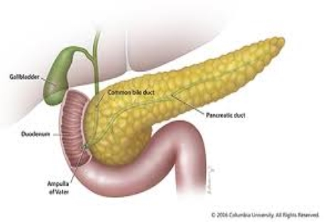
The Pancreas
Pancreas is a large gland which is both a digestive gland and an endocrine gland. It produces pancreatic juices which secretes along the pancreatic duct and this help break down food in the small intestine, along with other components such as bile and other fluids that helps metabolism of fats and proteins. This also consists of group of cells called the islets of Langerhans which make up the endocrine parts of the organ and produce hormones such as insulin and glucagon.
The pancreas is located below and behind the stomach, in the curve of the duodenum, which is part of the small intestine. This is a glandular organ that produces a vast number of hormones into the body and this form an integral part of the digestive system. This is an essential organ as it regulates glucose levels in the blood so if the pancreas stops producing insulin, it can lead to conditions like diabetes and a number of health issues.
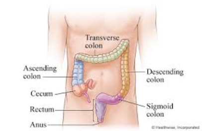
The large intestines
This is also known as a colon and is a thick tube that receives waste from the small intestine. It contains the caecum, colon, rectum and anal canal. It also consists of bacteria, which helps in breaking down any remaining food and provide more vitamins needed for the body. The colon contains four parts which are the descending colon, ascending colon, transverse colon and sigmoid colon.
The descending colon is the part of the large intestine which passes downwards on the left side of the abdomen towards the rectum whereas the ascending colon comes in the main part of the large intestine, which passes from the caecum on the right side of the abdomen. Transverse colon is in the middle part of the large intestine which passes across the abdomen from right to left below the stomach. Lastly, the sigmoid colon is the S-shaped which comes at the last part of the large intestine, leading into the rectum.
The water in the food we consume passes through the colon walls into nearby blood vessels. This plays in a much smaller role such as storing waste, reclaiming water, maintain water balance and absorb certain vitamins. As a result, it creates waste (faeces) which the body pushes out through the rectum, anal canal and anus (a hole surrounded by a muscular ring). The colon’s role in the body is to get rid of water, some nutrients and electrolytes from the digested food. The colon is the area that exchanges liquid to solids and then transported to the rectum.
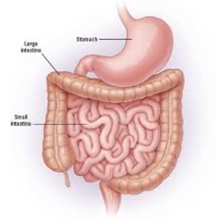
The small intestines
The small intestine has the main role in digestion which looks like a coiled tube with three parts- duodenum, jejunum and ileum. This also links up with the oesphagus, large intestine, and the stomach which forms the gastrointestinal tract. The internal walls of the small intestine consist of many tiny finger-like tissues called villi which all contains capillaries in which most of all the food gets absorbed. Each and every one of the villi is covered in even smaller finger-like structures called microvilli in which both villi and microvilli help increase surface area for the absorption of nutrients A lymph vessel called a lacteal absorbs recombined fat particles. Any remaining semi-liquid waste mixture passes into the large intestines.
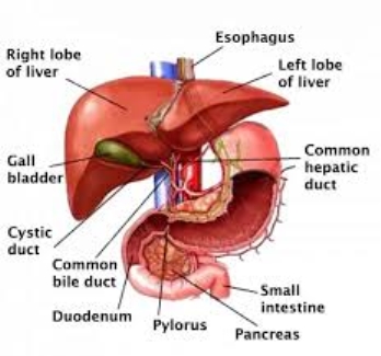
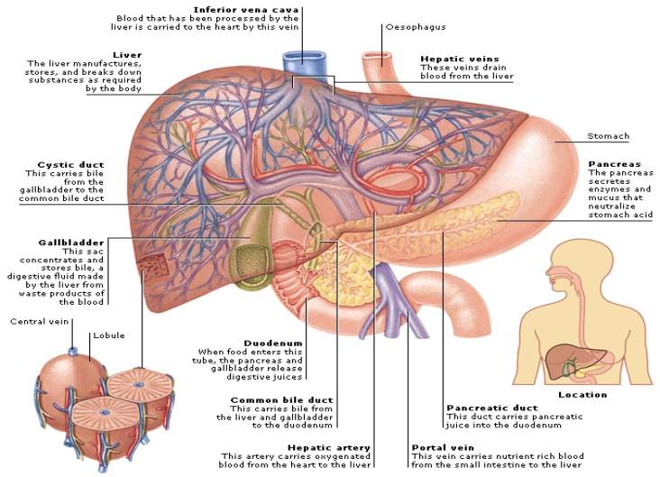
The liver
This is the largest organ which is located in the upper right portion of the abdominal cavity, under the diaphragm and to the right of the stomach, the liver contains four lobes. The average adult liver weighs about three pounds.
One of the many roles this organ plays in consists of being a digestive gland which secretes bile along the hepatic duct. One other role is it converts and store newly- digested food matter which comes from the hepatic portal vein. Overall, this help regulates the level of blood glucose and in destroying old red blood cells. Also, stores vitamins and iron and creates important blood proteins.
This is a vital organ as it supports nearly every organ in the body so without this, humans won’t be able to survive. It is known as a gland because it secretes chemicals such as bile- a substance which digest fats. Bile’s has salts which breaks down fat into smaller pieces so it is easier to get absorbed in the small intestine.
In addition to the production of bile, comes the liver. This detoxifies the blood by getting rid of harmful substances such as alcohol and drugs. And also, breaks down hemoglobin as well as insulin and other hormones. It converts ammonia to urea which is vital in metabolism.
![]()
The ovaries
This is a ductless reproductive gland in which the female reproductive cells are produced. It is held in place in the lower abdomen by ligaments. These helps attach them to the pelvic walls. The female sex cells called ova are being produced regularly in the ovaries right after puberty. An ovary is normally firm and smooth and is about the size of an almond.
The female reproductive systems consist of a vulva, ovarian follicles uterus and the vagina. The vulva comes in the outer parts of the female reproductive system including the labia and the clitoris. The labia are two folds of skin which surrounds the openings of the vagina and the urethra. The clitoris is the most sensitive part and like the penis, it is made of erectile tissue which has many receptors. The ovarian follicles are areas of tissue which appears regularly in the ovaries after puberty hits. Each has a maturing ovum and the follicles gradually increase in size which then secretes hormones. The uterus consists of the making of the baby. Lastly, the vagina is a muscular canal leading from the uterus out of the body. It carries the ova and endometrium during menstruation. It has a lining of mucous membrane covering a muscular wall with a vast number of blood vessels.
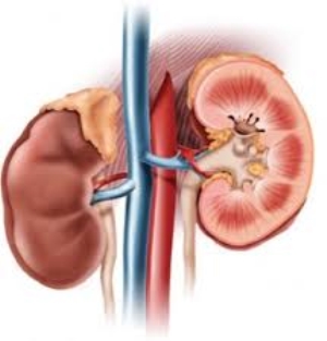

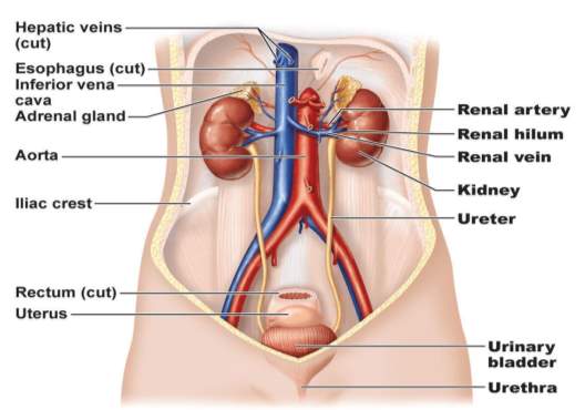
The kidneys
These are two bean-shaped organs that extract any waste from blood; balances body fluids, form urine and help other important functions of the body. They are located against the back muscles in the upper abdominal cavity and sit opposite each other on either side of the spine. The right kidney sits a little bit lower than the left to accommodate the liver.
The urinary system is the main system of body parts involved in excretion, which gets rid of unwanted substances. Kidneys are the main two organs in excretion filter out unwanted substances from the blood and regulate the level of body fluids. Blood enters through the renal artery and leaves it in renal vein. Furthermore, there are many things the body doesn’t need inside which the kidneys filter out such as excess salts, and urea, a nitrogen- based waste created by cell metabolism. Urea is synthesised in the liver which transfers through the blood to the kidneys to get removed.
More on the core actions of the kidneys include in balancing the level of water. As kidney’s role is to break down chemicals from urine, they react to any changes in the body’s water level throughout the day. When the water level decreases, the kidneys adjust accordingly and leave water in the body instead of helping excrete it. This also regulates the blood pressure because kidneys need constant pressure in order to filter the blood so when it drops too low, the kidneys increase the pressure. A way it does this is by producing a blood vessel constricting protein which signals the body to retain sodium and water. Both constriction and retention help restore normal blood pressure.
The kidneys also help regulate red blood cells so when the kidneys have a lack of oxygen, it sends a message in the form of erythropoietin, a hormone that stimulates the bone marrow to produce more oxygen-carrying red blood cells. Lastly, it regulates acid so as cells metabolises, it produces acids. The food that we consume can either increase the acid in our body or neutralises it. The body needs a stable balance of chemicals 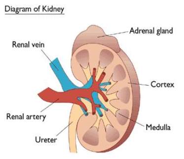 in order for it to function properly which the kidneys also do.


They are endocrine glands which produce a vast number of hormones including adrenaline and the steroids aldosterone and cortisol.

A part of the kidney where ultrafiltration occurs
Nnnnnnnnnnnn It is the innermost part of the kidney
 The two tubes which carries urine from the kidneys to the bladder
It carries blood from the heart to the kidneys
They are veins that drain the kidney and also carry filtered blood
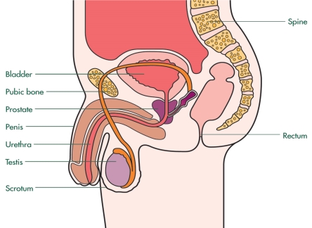
The testes
They are the most vital organs of the male reproductive system. They are glands where sperm and testosterone are produced. The testes lie in a sac which is located below the abdomen. They are contained in the scrotum and have a vast variety of dense connective tissue which contains around three hundred internal compartments called lobules. Each of these lobules contain a number of colled, tiny tubules where the sperm are produced. Testosterone is produced in cells located in between the lobules.
The penis is the organ through which sperm are ejected during sexual intercourse. It is made of soft, sponge-like erectile tissue, which has many spaces, blood vessels and nerve fibre endings. When a man gets sexually excited, the sinuses and blood vessels fill with blood making it stiff and erect.
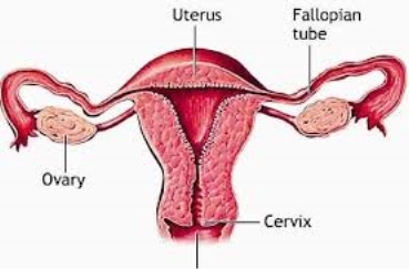
The Uterus
This is a hollow organ which is located inside, between the bladder and the rectum, in the pelvic area. Its role is to help develop the foetus and nourish it prior to birth. During menstruation, women releases eggs that travels through the fallopian tubes to the uterus. If fertilised, the eggs will bind themselves to the wall of the uterus in which the foetus can be developed. The uterus help nourishes and protect the foetus until birth.
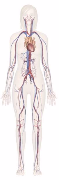
The Circulatory System
The circulatory system is a network of blood vessels, of which there are three main parts which are the arteries, veins and capillaries. Endothelium is a thin tissue layer which lines the arteries and veins and is the only layer of capillary walls. Blood continuously flow one way which is by the pumping of the heart, by muscles in artery and vein walls and by a decrease in pressure through the system.
The heart, blood vessels and blood itself are the three most vital components our body needs to survive. The system consists of two circuits in which blood can travel through and these are pulmonary and systemic. Activities such as exercises can affect the systems which cause the heart to pump blood faster around the body which as a result you can exercise for longer.
The circulatory system carries blood and of any dissolved substances to and from different places in the body. The heart has a role of pumping these things around the body and it pumps blood and substances through tubes called blood vessels. The heart and blood vessels work together which makes up the circulatory system.
The system is a double circulatory system which means it has two parts. The pulmonary circuit carries blood to the lungs to get renewed (oxygenated) and then back to the heart. In the lungs, carbon dioxide gets removed out of the blood and exchanges it into oxygen by the haemoglobin which is found in red blood cells. However, the systemic circuit carries blood around the body to deliver oxygen and gives back de-oxygenated blood to the heart. Blood can also carry nutrients and waste.
The heart contains veins which brings blood from the body but the lungs. The arteries in the heart carry blood away from the arteries whereas the coronary arteries supply its own blood. The heart is a muscular pumps so when it beats, it pumps the blood to the lungs and around the body.
The heart has four chambers which consist of two atria and two ventricles. Blood enters the heart through the two atria and exits through the two ventricles. There are valves which prevent the blood from flowing in the wrong direction and the septum separates the two sides of the heart.
There are three types of blood vessels in the body which are the arteries, veins and capillaries. The arteries carry blood away from the heart, have a thick muscular wall and have a small passageway for blood which also contains blood under high pressure. The veins can carry blood to the heart, have thing walls, contain blood under low pressure and have valves to prevent blood flowing backwards. The capillaries are found in the lungs and muscles, are microscopic and very low blood pressure. This is also where gas exchange takes place so when oxygen passes through the capillary wall and into the tissues, carbon dioxide passes from the tissues into the blood.
Our blood is essential for the body which consists of four key components which are plasma, red and white blood cells and platelets. An average adult can hold up about 5.5 litres which travels around in the circulatory system. The blood distributes heat and carries many more important components in its plasma. The old blood gets replaced by new ones through the process called haemopoiesis.
Plasmas are pale liquid which contains the red blood cells and it carries dissolved food for the body cells, waste matter and carbon dioxide are secreted by them, antibodies which combat infection and enzymes and hormones help with the body process. Red corpuscles other known as red blood cells are a red, disc-shaped without a nucleus. They are created in the bone marrow which also contains haemoglobin which gives it its dark red colour. When this is mixed with oxygen in the lungs, it forms oxyhaemoglobin which makes the blood darker. The red cells pass the oxygen to body cells by diffusion and then return back to the lungs with haemoglobin. The white blood cells are large and are an important part of the immune system which produces antibodies and kill of any harmful microorganisms. Platelets are very small disc-shaped bodies which are also made in the bone marrow. These clumps together to form clot and protect the body by stopping blood lost. It transports nutrients and waste, delivers oxygen to the working muscles and removes heat.
The Respiratory System
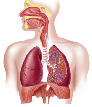
The human respiratory system is split into two parts, one being the lower and the other being the upper respiratory tract. When you breathe in, the air is already filtered through natural lines of defence which protects the respiratory tract from any illnesses or irritation. Nasal hairs in nose are there to protect any large particles of dust that might get inhaled the respiratory and so are the rest of the system is lined with mucous membrane which secretes mucous. The mucous help trap any small particles such as pollen or smoke. Cilia are like hairs and these lines the mucous membrane and move the particles that were trapped in the mucus out of the nose.
The air we inhale is moistened, warmed and cleansed by the nasal epithelium which covers the conchae in the nasal cavity. This has increased blood flow which helps warm the inhaled air, but also facilitates nosebleeds for some.
The pharynx is a muscular, funnel- shaped tube around 5 inches long which connects the nasal and oral cavities to the larynx. The pharynx protects the tonsils and the adenoids, which are lymphatic tissues that prevent infections by releasing white blood cells. The larynx creates the entrance to the lower respiratory system. The epiglottis is a leaf-shaped flap. This help prevents food or liquid from crossing the line when swallowing and there are two pairs of strong connective tissues which are wrapped around the vocal cords which vibrates making noise when talking or singing.
The lower respiratory tract consists of the windpipe and within the lungs, bronchi, bronchioles and alveoli. After the inhaled air transfers through the larynx, it reaches the trachea. It is a rigid tube about 4.5 inches long and 1 inch wide. This is located in the walls of the trachea and its distinct shape gives it its rigid look so that it stays open at all times. Inward airflow from the trachea then branches off to two bronchi. One leads to the right lung, while the other one leads to the left lung. This contains a C-shaped cartilage rings just the like the trachea.
In the lungs, there are bronchi that are split into two which are the secondary and tertiary bronchi which have a continuous line to the airways called the bronchioles. There is no cartilage in the bronchioles therefore they can be constricted and obstruct, as during an asthma attack. The end of the bronchioles ends with alveoli which are bundled together to form alveolar sacs. There is a connection of capillaries on each of the alveolus which carries blood through veins from other parts of the body. The gas exchange occurs here in which exchanges carbon dioxide to oxygen from the alveoli. After this has occurred, it goes to the heart which is pumped out to all body tissues and extremities. The carbon dioxide is then exhaled and expelled from the body when we breathe out.
The Nervous System
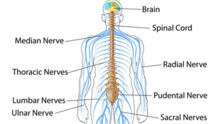
The nervous system consists of the central nervous system which contains the brain and spinal cord and the peripheral nervous system consisting of nerve cells that carries information to and from the central nervous system. Nerve cells are also called neurons and each one of them contains a cell body, an axon and one or more dendrites. There are three forms of neurons which are sensory, association and motor neurons.
Neurons have long fibre axons which is covered by a fatty sheath and tiny branches named dendrons which branch further as dendrites at each end. The cerebrum is the largest part of the brain which contains two hemispheres. This controls movement, speech, intelligence, memory, emotion and sensory processing.
At the base of the brain, the stem is connected to the spinal cord which is made up of midbrain, pons and medulla oblongata. The midbrain acts like a complex switchboard which allows the brain to communicate with the rest of the nervous system. The pons sends messages from the cerebrum to the cerebellum and spinal cord. Lastly, the medulla is the portion of the brain stem located just above the spinal cord. It plays a vital role in the body such as heartbeat and breathing.
The brain controls every function in the body and the nervous system gives of a message to the body in a way of responding to stimulus. Receptors detect any environmental changes which send impulses along the motor neurons to the effectors, which bring about a response.
The thalamus is in the central part of the brain which helps process and coordinates sensory messages like touch, received from the body. Hypothalamus regulates functions such as thirst, appetite and sleeping patterns. It plays in the role of releasing hormones from the pituitary gland. Pituitary glands are tiny and they produce hormones involved in growth, puberty, metabolism, water and mineral balance, the body’s response to stress and more. Cerebellum helps in coordination and the spinal cord runs down the inside of the spinal column which connects the brain with nerves going to the rest of the body.
The function of nerves in the nervous system is the autonomic nervous system which regulates certain body processes such as blood pressure and the rate of breathing. The somatic system consists of nerves that connect the brain and spinal cord with muscles and sensory receptors in the skin.
Sense organs consist of receptors that are sensitive to stimuli. There are five sense organs which are skin, tongue, nose, eyes and ears. They all are sensitive to different receptors such as skin being sensitive to touch, pressure, pain and temperature. Tongue is sensitive to chemicals in food whereas nose is to chemicals in the air. Eyes are sensitive to light and ears are sensitive to sound and position of the head.
The renal system
This system includes organs such as kidneys, where the urine is being produced, and the ureters, bladder and urethra for the passage, storage and voiding of urine. This is located in the pelvis which is held in place by ligaments attached to other organs and the pelvic bones.
The kidneys are two organs found along the posterior muscular wall of the abdominal cavity. The left kidney is slightly larger than the right kidney, making it is less superior than the left one. The kidneys are bean-shaped organs and are approximately 11.5 cm. Each kidney consists of millions of nephrons and each one of these are made up of a very small filter called glomerulus which is attached to a tubule. As the blood passes through the nephron, fluid and waste products gets filtered out.
The body absorbs all the nutrients from the food we consume in which gets used to maintain all bodily functions including energy and self-repair. Once the nutrients have been absorbed, waste products are left in the blood and in the bowel. The renal system works with the lungs, skin and intestines. All of these organs excrete waste which helps keep chemicals and water in the body balanced.
The renal system removes urea out of the blood which is a waste. This is produced when protein such as meat, poultry and certain vegetables gets broken down in the body. Urea gets carried around in our bloodstream to the kidneys. From the kidneys, urine travels through a thin tube called the ureters to the bladder. This is about 8-10 inches long and it contains muscles which help tighten and relax to force the urine downward away from the kidneys.
Sphincters are circular muscles which stops pee from leaking so these close tightly like a rubber band around the opening of the bladder to the urethra and this tube allows urine to pass outside the body.
The digestive system
The digestive system is a complex series of organs and glands in which help process food. This also helps break down food that we consume and use it as energy. This begins in the mouth by chewing and ends down to the small intestine. Once the food passes through the gastrointestinal tract, it gets mixed with digestive juices which help breakdown large molecules into small molecules. This gets absorbed by the body through the walls of small intestine into the bloodstream which gets delivered around the body. The waste then goes through the large intestine and out of the body as solid called stool which comes out from the rectum.
In the mouth, there are salivary glands which is a lubricating fluid consisting of enzymes that breakdown carbohydrates then the food goes pass the oesophagus which gets transported to the stomach. The chemicals in the stomach like the acid and enzymes breaks down the material through muscular contractions. The small intestine consists of enzymatic digestion and absorption of water, organic substrates, vitamins and ions whereas large intestines deal with dehydration and compaction of indigestible materials in order for elimination.
Furthermore, the digestive system is vital to the body as it gives us the energy to continue the tasks we do on the everyday basis or else we’ll die. This also helps eliminate waste that the body no longer need.
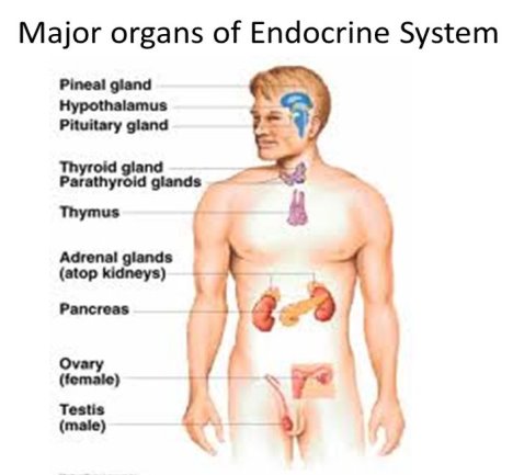
The Endocrine System
The endocrine system helps regulate vital functions in the body such as metabolism, growth and development, mood, maintain homeostasis and other more. The glands in endocrine system are controlled by direct stimulus from the nervous system. Glands are an organ in the body which secretes particular chemicals for the body to use or for discharge around the body. However, hormones are a regulatory substance and are produced in organisms which get transported in tissue fluid like blood, sap to activate specific cells or tissues to get going.
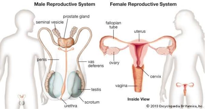
The reproductive system
The way both gender’s reproductive systems work is when the woman’s egg needs sperm in order to get fertilised. Out of the five thousand sperm that entered the body, only one can be fertilised.
The woman goes through menstrual cycle once a month which means an egg is released from the ovaries. During this period, the uterus prepares itself for the arrival of the embryo. It’s more than likely that the woman will not go through menstruation when they are pregnant which happens once the egg has been fertilised. Reproductive system’s role in a woman is to produce an egg and nourish it until birth.
The female reproductive system and endocrine system work together as the menstrual cycle is controlled by hormones which are of the endocrine system. The hormones will also start the process of the menstruation in women.
The male reproductive system’s role is to produce sperm to help fertilise an egg. Semen is a mixture of sperm and seminal fluid which comes between 50 million and 130 million in 1 ml of semen. A mature sperm cell’s structure consists of a head which contains a highly condensed nucleus and a tail or flagellum which propels the cell forward. A vast number of mitochondria are needed in order to get to the egg so when the sperm reaches the egg, enzymes in the sperm’s head can breakdown the egg’s outer layer. When a guy is sexually aroused, the automatic nervous system prepares to male organs to deliver sperm. The penis becomes erect, then sperm ejects from it by the contractions of smooth muscles lining the glands in the reproductive tract.
The musculoskeletal system
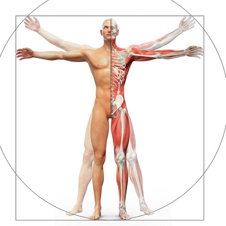
The muscular system consists of the muscles, bones, joints and ligaments. The muscles are responsible for body movements due to contractions of the muscles which is because of the movement of microfilaments. It provides movement along with maintaining posture, generating heat and stabilises the joints.
The skeletal system provides an internal framework for the body, which help protect organs by enclosure, and holds skeletal muscles in place so that muscles can move around. The bones also provide protection to any soft organs in the body which could get damaged easily if there were no protection such as bones to protect them. They also store fat in the internal cavities which help store minerals.
Skeletal muscles are attached to bone by tendons in which is being used as levers in order for the body and its parts to move. As the muscles increase in size, the bones must also do the same to support the bigger muscles or else it would break. There must be calcium in the blood so that the nervous system can send messages to muscles to move and for blood to clot when your skin gets injured. Calcium is found in bones which helps provide movement for the body from the muscular system.
There is a total of 206 bones in the human body and these are situated with precision, in locations to provide structure and support. There are 22 bones separated that are fused together to provide protection for the brain, eyes and ears.
The upper limb consists of humerus, radius and ulna. The humerus is connected to the thorax and the lower arm is made up of two bones like the radius which is thicker and ulna the thinner one.
Ligaments are a strong band of tissue which holds the ends of bones together at a joint which is where two or more bones meet together. Tendons are cords of connective tissues which help attach muscle to bone.
There six main kinds of joints which are ball and socket, hinge, gliding, saddle, pivot and fixed joints. The ball and socket joint forms when the round head of one bone fits into the cavity of another. A hinge joint provides a flexing and extension movement, just like how door hinge work. A saddle joint forms when the surfaces of both bones joined have a concave and a convex region. The fixed joint is joints found in the skull which does not move at all.
The digestive system contains many organs that work together in converting food into energy along with giving the body the right nutrients. As food gets eaten, it goes through a long tube inside the body called the gastrointestinal tract. This consists of the oral cavity, pharynx, oesphagus, stomach, small intestines and large intestines. In addition to the gastrointestinal tract, there are vast numbers of other organs which help with the process of digestion.
The structures that are responsible for ingestion, chewing, swallowing and initial digestion of food are of the digestive system of the head and neck. It includes the mouth, salivary glands which produces saliva and of the pharynx. Ingestion of food begins the process of digesting food which is through the mouth. Once the food has entered the mouth, we start to masticate so that large masses of food can be broken down in order to get through the gastrointestinal tract. The breakdown of food can also be known as mechanical digestion which is assisted by the tongue, lips and cheeks. These help by moving the food around the mouth in order to get chewed up by the teeth.
There are three salivary glands surrounding the mouth which all secrete saliva into the oral cavity. The first gland is called the parotid glands which can be found on either side of the jaw just anterior to the ears. This gland secretes saliva into the posterior of the mouth. The second gland is the submandibular glands which are found just anterior and slightly inferior to the parotid glands. These are found just under the jaw and they secrete saliva into the middle part of the mouth. Lastly, the third glands are called the sublingual glands which secrete saliva into the anterior part of the mouth and can be found under the tongue.
The saliva is mainly made of water, but also contains enzyme such as salivary amylase and lingual lipase. The saliva helps moisten and soften dry food in the mouth so that delicate mucosa of the digestive system can be protected as this also help with the process of swallowing food. The process first begins with the salivary amylase through chemically digesting carbohydrates. Starches from food are broken down into simple sugars that can be used as a fast energy source. Lingual lipase works the same way too as it converts fats into fatty acids, but this only starts up when food reaches the acidic environment of the stomach.
A bolus is chewed up food containing a mixture of saliva which forms a paste-like substance rolled into a mass. This mass goes down the pharynx through a complex interaction of muscles of the mouth, tongue, palate and throat. The swallowed food first goes through the pharynx which is funnel-shaped then passes through the oropharynx region at the back of the mouth and then through the laryngopharynx that connects to the oesphagus and larynx. Epiglottis is a flexible fibrocartilage which is found just above the larynx and this is important as it prevents masses of food going down the wrong hole. The movement caused by the epiglottis is also vital as it stops food from blocking the airway which could lead to serious conditions such as asphyxiation. The bolus of food goes through the oesphagus, which is where it will get directed to the stomach by muscle movement known as peristalsis.
The gastrointestinal tract is the upper abdomen and chest area of the digestive system. This consists of many important organs such as oesphagus, stomach, duodenum, liver and gallbladder. These organs all contribute in breaking down masses of food and direct them through the alimentary canal.
Chewed up food enters through this long, tube-like oesphagus from the throat which is helped by peristalsis for movement towards the stomach. When the food has gone down to the stomach, the acid and digestive enzymes activates and forms chyme which converts solid into liquid. Once chyme has been produced, it goes through the duodenum which is the first segment of the small intestine. The liver and gallbladder produces bile along with enzymes from the pancreas gets mixed with the chyme in the duodenum in order to breakdown more nutrients from the food in which the intestines absorb.
The process of the chewed-up food then goes down to the digestive organs of the lower torso. This includes lower part of the gastrointestinal tract which is the small intestines, large intestines and the rectum. The liver and pancreas both assist the gastrointestinal tract in digesting nutrients out of the food. When the food has reached the end of the gastrointestinal tract through the rectum, it means all of the nutrients from the food have been sucked out of it. Bacteria then help with the digestion of food and convert this into feces while absorbing the water out of it so that only solid soluble can be excreted.
The chyme reaches the lower part of the GI tract through the pyloric sphincter of the stomach. Chyme goes through this tube called pyloric sphincter then enters the duodenum which is a foot long of C-shaped segment of the small intestine which is located just inferior to and to the right of the stomach. Furthermore, the duodenum also receives bile from the gallbladder and liver, along with pancreatic juice. Both pancreatic juice and bile gets mixed with the chyme in the duodenum so that the acid from the chyme can be neutralised, emulsify lipids, and chemically digest the chyme into its most basic building blocks.
The walls of the duodenum also absorb small amounts of nutrients from the food but most of it gets absorbed in the jejunum. Jejunum is eight-foot-long and belongs in the middle part of the small intestine which is located between the duodenum and ileum. Once the chyme has entered the jejunum, it means it has been thoroughly digested and now goes through the process of getting all the nutrients absorbed through the intestinal mucosa. The mucosa of the jejunum consists of tiny villi along with vast numbers of circular folds along its length. These help by increasing the surface area of the jejunum which allows it to absorb more nutrients. Once the chyme has gone pass the jejunum, it enters through the ilium which 90% of the nutrients get absorbed into the bloodstream.
Chyme goes through till the end of the small intestine to the ileum which is a 10-foot long tube which leads to the cecum of the large intestines. The chyme enters the ileum with very little nutrients and these still gets absorbed by the ileum before making its way down to the large intestine. The ileum also has villi and folds the same as the jejunum so that surface area can be increased in size in order to take in more nutrients. Peyer’s patches also known as lymphatic tissue lines the ileum and keeps an eye for pathogens entering the intestines so that any illnesses can be prevented.
Once it has reached the end of the ileum, the chyme goes through the ileocecal sphincter and enters the large intestine. The cecum is a dead-end pouch on the right inferior end of the large intestine. The chyme then gets mixed with bacterial flora which produces into feces. This then gets moved by peristalsis out of the cecum and into the ascending colon while bacteria start to break down indigestible waste material. Vermiform appendix is linked with the inferior end of the cecum which is a thin tube that stores beneficial bacteria. The materials then go through the colon which is the longest section of the large intestine in which extends from the cecum to the rectum. Colom has four major regions which are the ascending colon, transverses colon, descending colon and sigmoid colon. The ascending colon connects to the cecum and run superiorly towards the right inferior corner of the liver. Under the kidney is where you would find the ascending colon and to the left is the transverse colon. The transverse colon crossed to the left side of the abdomen, turns to the downward which will be the descending colon. The descending colon runs down the left side of the abdomen to the sigmoid colon. Lastly, the sigmoid colon is an S-shaped end to the colon that terminates in the rectum.
Waste materials passes through the large intestine gets mixed with and fermented by bacteria. These bacteria then release vitamins B and K from the waste material as they move along the colon. The walls of the colon then absorb the vitamins K and B along with water and any other left nutrients. Once the feces have reached the end of the colon, the waste materials are left of dried, condensed and no vitamins and nutrients in it.
The rectum is a short, downward tube near at the end of the large intestine which stores the waste materials until the body is ready for elimination. It is lined with vast number of sensory receptors which looks out for pressure and stretching in the walls of the rectum. When it has filled with waste, receptors send messages to the brain to let the conscious mind know that waste is ready to be eliminated. The anal canal is the last segment of the large intestine which is found at the end of the anus and this controls the discharge of waste.
Faeces that enters the rectum and anal canal applies pressure on the internal anal sphincter which causes the muscles to relax and dilate. The skeletal muscles of the external anal sphincter hold the waste in the anal canal until signals of dilation has been triggered voluntary. After both anal sphincters are opened, muscle contractions in the rectum and sigmoid colon push the waste through the anal canal and exeunt the body.
References:
- Colchester Institute Moodle (DEAD LINK)
- Colchester Institute Moodle (DEAD LINK)
- GCSE Science – BBC Bitesize
- Endocrine System (for Teens) | Nemours KidsHealth
- Reproductive and Endocrine Systems, Prezi
- How the Endocrine system & Nervous system work together, Prezi
- How does the organs of the digestive system work together, Prezi
- Introduction to the Digestive System – Organs and processes by which foodstuffs are broken-down by the human body.
- Human Renal Systemm, Brittanica
- Anatomy of the Urinary System – Stanford Medicine Children’s Health
- The structure and function of the nervous system – Coordination and control – The nervous system – OCR Gateway – GCSE Biology (Single Science) Revision
- Responding to changes in environment – How does the nervous system help us respond? – OCR 21st Century – GCSE Combined Science Revision
- Structure of blood and blood vessels – Cardiovascular system – Edexcel – GCSE Physical Education Revision – Edexcel – BBC Bitesize
- Interactive Guide to the Digestive System | Innerbody
Cite This Work
To export a reference to this article please select a referencing stye below:
Related Services
View all


DMCA / Removal Request
If you are the original writer of this assignment and no longer wish to have your work published on UKEssays.com then please click the following link to email our support team:
Request essay removal