Cut Mark Form and Deterioration in Heat Damaged Bones
Info: 9302 words (37 pages) Dissertation
Published: 16th Dec 2019
Tagged: PhysiologyMedicine
A R T I C L E I N F O A B S T R A C T
Cut marks on bone contain useful characteristics which are able to aid in the identification of the tool used as well as method of administration. There has been significant research into the implications of burning on bone and cut mark analysis as separate subjects. Understanding the influence of one upon the other is an area which still remains largely uncovered. This study presents information about the effect of direct heating on both pre- and post- burning cut marks.
Cut marks (n=16) were inflicted upon various animal bones (n=9) with an unbranded kitchen knife either prior to or following heat exposure. A temperature of 600°C was applied for a total of 3.5 hours by use of a muffle furnace. Any physical alterations to the cut marks were documented alongside the bone weight and total cut mark length.
Results indicate that cut marks added pre- and post- heat exposure exhibit a differing degree of colour change. Post- burning cut marks also display a greater degree of chipping at the point of exit of the knife. Although there was extensive fragmentation and heat fracturing across all bone samples, cut marks remained largely unaffected. In all cases aside from those where fragmentation had completely destroyed the bone structure, cut marks could be identified following the full length of heat exposure.

Keywords:
Cremation
Heat fractures
Cut mark characteristics
Burnt bone
Sharp force trauma
Fragmentation

1. Introduction
Disposing of a body by means of burning is not an uncommon occurrence during a criminal investigation. With the first legal use of cremation in the United Kingdom being in 1885 (Serpell,. 2009), the concepts and theories behind heat treatment as a means of disposal had existed for decades before they were adopted by criminals. Perpetrators of homicide will often go to great lengths in order to conceal their activity, with disposal of the body being of paramount importance. Cremation of a corpse is one such method of doing so, however, a less common practice is that of dismemberment. Dismemberment of a corpse may lead to cut marks presenting on the bones (Flieger et al., 2016). Abundant research has been carried out on these two methods in isolation (e.g. Humphrey and Hutchinson, 2001; Saville et al., 2007; Shipman et al., 1984; Thompson, 2004). however, little information is available regarding these techniques in conjunction with one another. Differentiating between cut marks which were produced prior to or following cremation could be an integral part of piecing together the events leading up to an individual’s death.
When bone undergoes heat treatment there are various observable characteristics which present as the bone is continually heated. External factors such as temperature, the time heat is applied, relative humidity and oxygen levels alongside internal factors such as bone composition, the amount of soft tissue present and age of the bone will define how it reacts (Waltenberger and Schutkowski, 2017). The differences within these categories will determine the extent of heat fracturing, weight loss, shrinkage and deformation of the bone during heat treatment (Waltenberger and Schutkowski, 2017). When exposed to high temperatures, the organic components of the bone are removed, followed by reestablishment of the inorganic materials. (Thompson, 2005). This has been found to lead to mechanical failure and fracturing within the bone(Waterhouse, 2013),alongside splitting and bone warping (Robbins et al., 2015). The effect of the heat may be lessened if there is soft tissue present, other protective material preventing direct heating of the bone such as clothing, or a limited oxygen supply. The four major stages in the process of cremation have been summarised as dehydration, subsequent decomposition, whereby organic components are removed, inversion; the removal of carbonates, and finally fusion, at which point the crystals melt (Mayne Correia, 1997). These stages were later reevaluated by Thompson in 2004 based on more recent literature and experimentation.
In cases which involve the explanation of observable trauma found on skeletal remains, elements of the cremation process may be inhibitory. Heat related fracture lines may act to obscure ante-mortem preexisting trauma, as can fragmentation (Ubelaker, 2009). However, several studies have documented the survival of preexisting trauma following heat exposure (Pope and Smith, 2004). It has also been noted that cut marks can be identified in bone which has been cremated, but their appearance may be altered by fragmentation following heat exposure (de Gruchy and Rogers, 2002). It is achievable to differentiate heat-induced fracturing from traumatic fracturing (Herrmann and Bennett, 1999), although there is insufficient research stating which particular characteristics of a cut mark are able to survive the burning process.
When a cut mark is produced on a bone surface there are two regions of forensic significance, these being the kerf wall and the kerf floor (Robbins, 2015). The kerf wall provides the most information about the particular saw or knife which created them (Freas, 2010), with the kerf floor yielding information about the saw form by displaying the relationship of saw teeth to one another (Symes et al., 2010)
Dismemberment is the method by which the limbs of a body are severed and the body cut into smaller fragments (Dogan et al., 2010). This is a factor which may lead to cut marks presenting on the bone which require identification. Ritualistic dismemberment may be sacrificial as was once common practice in Mesoamerica (Chinchilla Mazariegos, 2014). It has also been used as a means of capital punishment or during the process of cannibalism (Perez, 2012). In terms of criminal activity dismemberment is again a means of disposal of a body, making it easier for transportation. It can also be used to obscure or hinder positive identification of remains or finally, symbolically, as an expression of complete disregard towards the human body. (Symes et al., 2002).
Of the research conducted to date, there is little evidence of a comparative study between pre- and post- burning cut marks and their individual characteristics. It must be established whether it is possible to differentiate between these. The effect that localized direct heating has on cut mark presentation and deterioration must be demonstrated. Furthermore, the implications these two concepts could have towards the fields of Forensic Science, Forensic Anthropology and criminal applications requires investigation.
2. Material & Methods
Various bone types were sourced from multiple places for use within this study. In order to establish a protocol which could be followed throughout the duration of the study, preliminary testing was conducted upon cow (Bos) marrow bones sourced from a supermarket. A lamb (Ovis) femur sourced from a butcher was also used during this stage. The preliminary testing allowed any errors or lack of a clear protocol to be eradicated before the main experimentation was conducted. Animal bones were used in place of human bones due to ethical considerations. Research on decomposition and trauma cannot be done with cadavers in the United Kingdom without a licence to do so. Animals are often used instead, with pigs being the common choice as a human analogue within forensic experimentation (Lynn and Fairgrieve, 2009; Waterhouse, 2013a; Waterhouse 2013b;). It should be noted, however, that the mineral density is considerably lower in pig bones than in humans (Aerssens et al., 1998; Prat et al., 2012).
To investigate the effect of heat on both pre- and post- incineration cut marks, semi-fleshed pig (Sus) and lamb bones were cut and later heated. In this instance semi-fleshed refers to the removal of the majority of the overlying muscle tissue, with some underlying tissue and periosteum still present. Multiple different bone types were used within the main part of the study, firstly, pig foot bones comprising of metacarpals, first phalanx and an articulated section of the foot which were sourced from a supermarket in the form of trotters and subsequently partially defleshed. A lamb scapula, humerus and semi-fused radius and ulna were also tested. Due to the juvenile state of the bones, supported by the fact the epiphyses were not fully fused, it can be suggested that the bones are likely to have contained a higher amount of organic material. They will also have been less mineralized than those of an adult animal (Morgan and Bouxsein, 2008).
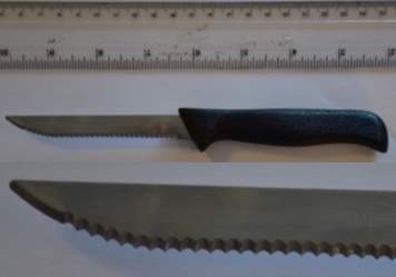 Each bone was measured according to guidelines set out by Angela von den Driesch in 1976 and the resulting measurements noted. Part of the radius bone had been cut off during butchering and so therefore only partial measurements of the radius could be taken for this bone. Both an osteometric board and digital sliding calipers (Mitutoyo absolute digimatic) were used to take the measurements. The bones were then weighed using a ‘Ohaus Pioneer’ two place balance. Once weighed cut marks were made in some of the bones using an unbranded kitchen knife. The knife had a total length of 193 mm, a blade length of 97 mm and a weight of 24.14 g respectively. The blade height measured 11 mm at the maximum with a serrated cutting edge, the spine of the knife is straight along the entire length. There is a space of 2.5 mm between each of the “teeth” that form the serrated cutting edge. An image of the knife used within the study can be found as Figure 1 below.
(a)
(b)
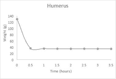
Fig. 1. (a) The knife which was used throughout the study to create the cut marks (b) Detail of the serrated cutting edge.
(b)
 Photographs were taken before and after the addition of the cut marks and following each stage of the study. After the cut marks were inscribed onto to particular bones, they were placed into crucibles and then into a ‘Phillip Harris Ltd. CSF 1200’ furnace set to a temperature of 600 °C. The preliminary testing allowed a suitable temperature to be selected as if too high this would not allow suitable changes to be identified following each stage, and too low would leave a large time frame before observable changes began. The bones were placed inside the cold furnace and heated. Once the temperature reached 600 °C they remained in the furnace for half an hour, this process was repeated seven times giving a total burn time of 3.5 hours. This was considered as sufficient to burn most organic material as Bohnert et al., 1998 found that temperatures of 670°C – 810°C will lead to complete incineration of the human body after approximately 2-3 hours. Following the half hour intervals, the furnace was switched off and the samples allowed to cool to room temperature. The length of the cut marks were re-measured after each interval and photographed, any changes were also noted at these stages. Additional post- burning cut marks were added and measured prior to the beginning of the next heating interval.
(c)
3. Results
The initial measurements and weights of each bone used within the study have been included below as Table 1.
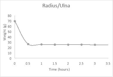
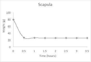
Fig. 2. (a) Total weight loss exhibited over 3.5 hours in Ovis scapula (b) Total weight loss exhibited over 3.5 hours in Ovis humerus (c) Total weight loss exhibited over 3.5 hours in partially fused Ovis radius and ulna.
Each tested bone exhibited weight loss beginning after the first half an hour session of exposure and continuing through to a plateux following approximately the third half an hour session i.e. 1.5 hours total heating time. Figure 2 shows the weight loss which three of the tested bones underwent across the 3.5 hour period for which they were tested.
(a)
| Initial Measurements (mm)a b c | ||||||||||||||
| Weight (g) | GL | Bp | Bd | Sd | Hs | DHA | SLC | GLP | LG | BG | BT | L1 | ||
| Preliminary Bonesd | ||||||||||||||
| Ovis Femur (P1) | 175 | 53.34 | 41.61 | 19.76 | ||||||||||
| Bos Marrow 1 (P2) | 123 | 48.34 | 48.18 | 41.35 | ||||||||||
| Bos Marrow 2 (P3) | 120.5 | 56.32 | 53.27 | |||||||||||
| Main Study Bones | ||||||||||||||
| Sus FourthMetacarpal (M1) | 24.89 | 78 | 21.57 | 23.07 | 17.45 | |||||||||
| Sus Fifth Metacarpal (M2) | 6.89 | 58 | 7.74 | 13.50 | 7.06 | |||||||||
| Sus Articulated Carpal Bones (M3) | 18.61 | |||||||||||||
| Sus First Phalanx (M4) | 17.94 | 51 | 18.04 | 17.43 | 17.67 | |||||||||
| Sus First Phalanx (M5) | 17.54 | 53 | 19.94 | 15.33 | 15.60 | |||||||||
| Sus Articulated Metacarpals (M6) | 76.66 | |||||||||||||
| Ovis Scapula (M7) | 79.38 | 149 | 157 | 26.18 | 41 | 31.33 | 26.57 | |||||||
| Ovis Humerus (M8) | 129.70 | 152 | 48 | 35.90 | 18.11 | 23.95 | ||||||||
| Ovis Radius/Ulna (M9) | 69.42 | 164 | 33.91 | 46 | ||||||||||
Table 1
Initial weights and measurements for the bones used within the study.
- Cases where a specific measurement could not be taken for certain bones have been indicated with a grey area.
- Abbreviations are as follows: GL = Greatest Length, Bp = Greatest breadth of the proximal end, Bd = Greatest breadth of the distal end, Sd = Smallest breadth of diaphysis. For scapula only: Hs = Height along the spine, DHA = Diagonal height, SLC = Smallest length of the Collum scapulae, GLP = Greatest length of the glenoid process, LG = Length of the glenoid cavity, BG = Breadth of the glenoid cavity. For Humerus only: BT = Breadth of the trochlea. For Ulna only: LO = Length of the olecranon
- Measurements were taken according to von den Driesch, 1976.
- Preliminary bones were not weighed, this was noted and used to assist in establishing the experimental protocol
Looking at the graphs it can be seen that following the first half an hour of heating there was a considerable loss of weight in each bone. The scapula, humerus and radius/ulna lost 66%, 72% and 62% of their total weight respectively following the initial heating phase.
In terms of overall weight loss, the percentage decrease following the full duration of heat exposure upon all bones has been compiled as Table 2 below. The values range from 64%-74% with the average percentage decrease being 71%. This shows a considerable weight loss across all tested bones following 3.5 hours heating at 600°C.
Table 2
Weight loss experienced by each bone over the duration of the study, showing percentage decrease.
| Bonea | Initial Weight(g) | End Weightb(g) | Percentage Decreasec(%) |
| M1 | 24.89 | 7.29 | 71 |
| M2 | 6.89 | 2.22 | 68 |
| M3 | 18.61 | 4.91 | 74 |
| M4 | 17.94 | 5.12 | 71 |
| M5 | 17.54 | 4.59 | 74 |
| M6 | 76.66 | 20.68 | 73 |
| M7 | 79.38 | 25.82 | 67 |
| M8 | 129.70 | 34.88 | 73 |
| M9 | 69.42 | 25.19 | 64 |
- For abbreviations please refer to Table 1
- End weight refers to the weight of the bone once all heating sessions were completed
- Percentage decrease example calculation for M1: 24.89-7.29 = 17.6, 17.6/24.89=0.707, 0.707*100 = 70.7 = 71%
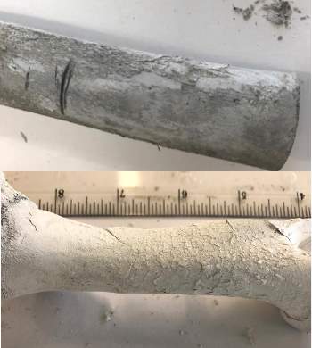
In terms of the total length of the cut marks which were placed into some of the bones, those which survived the heating process displayed considerable variation in the alterations they underwent. The lengths fluctuated, with varying degrees of increase and decrease occurring following the various heating stages. The vast majority of the cut marks were unmeasurable after the heating had begun due to heat fracturing and fragmentation greatly affecting their overall appearance. Many of the bones fractured along the cut marks, becoming fragmented as can be seen in the two Sus metacarpals which are pictured below as Figure 3.
(a)
Fig.3. (a) A metacarpal displaying a fracture line running down the length of the diaphysis which has distorted the cut mark form (b) A metacarpal displaying fragmentation across a cut mark resulting in complete separation.
The discoloration of the burnt bones ranged from the initial pre-burning pink-yellow all the way through to the resulting pure white. The samples became very fragile following the heat exposure with a chalky appearance to them. Nicholson, 1993, found that sheep bones burned in a muffle furnace at 600°C for 2.5 hours had predominant light grey discoloration. More recently in 2014 Imaizumi found that after burning for either 30 minutes or 2 hours, there was no time-dependent difference in the observed discoloration. Bones burnt at 500°C – 600°C appeared an ash-like gray colour following both of the allotted burning time periods.
The cut mark characteristics were largely unaffected by the heating process. The main observable differences between pre- and post- incineration cut marks was the degree of colour change which the cut itself underwent and the degree of chipping at the point of exit of the blade.
The visibility of those cut marks which survived the heating process remained clear, none of the effects of the heating process acted to obscure the marks other than those previously mentioned. In the instance of the radius bone a small mark which was produced when the knife slipped during an initial cutting stage remained visible following the full 3.5 hours of heat treatment. This can be seen in Figure 4.
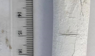

Fig. 4. Radius diaphysis with slip mark visible. The slip mark has been highlighted with a box.
As the heating progressed many of the bones exhibited a weathered texture to both the diaphyses and epiphyses. The epiphyses themselves had become detached early on during the heating stages, with most being fully detached by the third session, 1.5 hours total heating time. Flaking of the outer surface was visible among many of the bones. The outermost concentric thin layers of bone more commonly show evidence of flaking following subaerial weathering over a prolonged period of time (Behrensmeyer, 1978). Examples of the flaking which was observed during this study can be seen below as Figure 5. A dry and porous texture was seen across all bones following the completion of all seven heating sessions. Following detachment of the epiphyses from the diaphyses it was noted that many of the epiphyses underwent alterations consistent with subaerial weathering.
| Total Cut Mark Length (mm)b
Table 3 |
|||||||||
| Time (hours) | |||||||||
| 0 | 0.5 | 1 | 1.5 | 2 | 2.5 | 3 | 3.5 | ||
| Bonea | |||||||||
| M1(a) | 13.85 | 10.89 | 11.84 | 11.87 | 11.80 | 11.72 | 11.77 | 11.54 | |
| M1(b) | 14.28 | ||||||||
| M (c) | 11.25 | 11.06 | 11.47 | 11.47 | 11.48 | 11.40 | 11.36 | ||
| M1(d) | 12.63 | 12.55 | 12.61 | 12.35 | 12.49 | 12.35 | |||
| M2(a) | 4.99 | 5.35 | 5.20 | 5.06 | 4.89 | 4.86 | 5.11 | 5.32 | |
| M2(b) | 10.86 | 10.01 | 9.92 | 9.74 | 9.47 | 9.57 | 9.54 | 9.84 | |
| M3 | 14.59 | 15.17 | 14.51 | 13.84 | 14.43 | 13.56 | 12.30 | 14.80 | |
| M4 | 5.89 | 5.23 | 5.13 | 5.00 | 4.88 | 4.49 | 4.41 | 4.40 | |
| M5 | 8.50 | 8.51 | 8.25 | 8.19 | 7.94 | 6.13 | 6.10 | ||
| M7 | 29.51 | ||||||||
| M8 | 15.35 | 14.61 | 14.96 | ||||||
| M9(a) | 12.08 | 11.50 | 11.15 | 11.41 | 10.87 | 10.18 | 10.53 | 10.51 | |
| M9 (slip)c | 5.03 | 5.15 | 4.67 | 4.59 | 4.88 | 4.17 | 4.17 | 4.05 | |
| M9(b) | 11.22 | 10.75 | 10.72 | 10.33 | 10.55 | 10.61 | 10.69 | ||
| M9 (c) | 11.36 | 11.76 | 11.64 | 12.03 | 11.94 | 11.86 | |||
| M9(d) | 11.64 | 11.60 | 11.93 | 11.58 | 11.48 | ||||
(a)
(b)
Fig. 5. Weathered and flaking appearance to bones. (a) Radius following 0.5 hours total heat time (b) Humerus following 3.5 hours total heat time.
Due to the fragility experienced by the bones following heat exposure, shrinkage and deformation was not able to be well documented. Accurate measurement of the greatest length amongst others could not be retaken once the heating sessions had begun due to the likelihood of damage occurring to the bones whilst the measurements were being taken.
As previously mentioned total length of the cut marks was measured throughout the study where possible and these measurements have been compiled as Table 3. In many cases measurements were unable to be taken as the cut marks were either distorted or completely concealed by various heat related physical alterations. Interestingly, in the instance of the humerus bone a cut mark was still visible on a fragment of bone which had become detached (Figure 6)
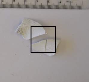
Fig. 6. Fragments of bone detached from the humerus. The box indicates where a cut mark is still visible across the fragments.
- For abbreviations please refer to Table 1.
- Instances where no measurement could be taken have been indicated with grey shading
N.B. where a grey area lies before other measurements this indicates the cut marks were added post- heat treatment
- Slip refers to the unintentional cut mark which occurred when the knife bounced during addition of cut mark M9 (a)
As can be seen within table 3, there were varying degrees of increase and decrease for the total cut mark length. The scapula (M7) and humerus (M8) very quickly became fragmented which meant the cut marks placed on them could not be measured following heat treatment.
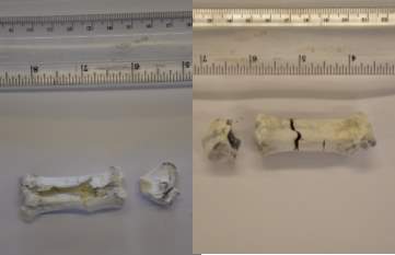
Both articulated and disarticulated metacarpals were placed in the furnace in order to establish whether there was a difference in the effects of heat treatment between them. Figure 7 below shows the fifth metacarpal of both the articulated and disarticulated metacarpals.
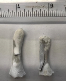
Fig. 7. Sus fifth metacarpals from articulated foot bones (right) and disarticulated foot bones (left) following 2.5 hours of heat exposure.
As can be seen in figure 7, the metacarpal which remained in articulated form prior to heat treatment shows increased fragmentation and a slower rate of colour change.
Extensive fragmentation occurred within the furnace, alongside heat fracturing. In accordance with Schmidt and Symes (2015) classification, the fractures were mainly longitudinal and step fractures, although a curved transverse fracture was noted in one instance. Figure 8 shows this fracture on the posterior side of the radius.
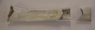
Fig.8. A curved transverse fracture of the radius shaft.
4. Discussion
When a bone is directly exposed to heat there are certain variables which undergo alteration as exposure continues at a predetermined rate. The interactions between these variables and both pre- and post- burning cut marks is largely unknown. The first variable to be considered in terms of heat alterations is that of colour change.
Coloration of burnt bones is a topic which is continually debated. Many studies have attempted to establish a relationship between bone colour and temperature (DeHaan and Nurbakhsh, 2001; Devlin and Hermann, 2015; Mayne Correia, 1997; Shipman et al., 1984; Stiner et al., 1995) in order to assist in estimating the exposure temperature of unknown cremated bone samples. There are multiple factors which lead to variation in this area including duration of heating, oxygen availability, amount of soft tissue present and sample size as well as type of furnace used (Imaizumi, 2015). The general order of discoloration begins with an unburnt tan/yellow appearance, moving through a sequence of brown to dark grey/black, blue and grey and then finally moving towards pure white. This is consistent with the stages of transformation that the bone is undergoing at the same time, the brown to black appearance emerges following the process of carbonization, whereby the organic materials of carbon and collagen are incinerated. This is superseded by decomposition of the organic compounds, leading to a grey discoloration. Finally, the emergence of a white colour is a culmination of a complete loss of all organic compounds within the bone and fusion of bone minerals (Ellingham et al., 2015). Although the sequence of colour change is constant, the temperatures attributed to them tend to vary between studies (Shipman et al., 1984; Quatrehomme et al., 1998). Nicholson, 1993 found that the onset of colour change varied notably between the bones of mammals, fish and birds. It has also been noted that bones may experience more than one colour stage at the same time, sometimes the whole range of colours can be noted on a single bone in the same instance (REFERENCE). Since within this study a muffle furnace was used, this would have allowed uniformity in temperature with each area of bone experiencing the same temperature. In terms of a domestic fire or other heat source, this may not be a constant. Certain areas of bone may receive a higher temperature at times by being in direct contact with a flame compared with other areas of the bone. This may be especially relevant in human bones, especially longer bones such as the femur where the bone may be experiencing multiple temperature fluctuations at the same time.
In terms of interpretation of colour it is likely that individuals will have slight variations within the way they perceive and describe colours. Shipman et al, 1984 were the first to employ the Munsell Soil Colour Chart (Munsell Color Company Inc, 1994) as a means of standardizing colour definitions. Within this study a visual assessment of the bones was sufficient as the main focus was the effects of heating upon the cut marks. Bones were found to go from an unburnt beige colour through to dark/grey black and eventually light grey/white. Time taken to reach a full white discoloration varied between bones, the Sus foot bones became white more rapidly than the scapula, humerus and radius/ulna. Since coloration is a reflection of both the organic and inorganic materials associated with the bones as they respond to the increased temperature (Mayne Correia, 1997) the differences could be associated with varying levels of these components within particular bone types.
The state of the bone prior to heat treatment could also have an effect on the rate of discoloration. As can be seen in figure 7, a metacarpal which was heated whilst still in articulated form experienced a slower rate of color change than the same bone in disarticulated form. This could be due to the other foot bones shielding the smaller bones from direct heat exposure, resulting in less discoloration after the same period of heating. It was also found that the diaphyses moved through the color stages more rapidly than the epiphyses. The epiphyses themselves became detached from the diaphyses fairly rapidly after heat treatment had started. This could be a reflection of the juvenile nature of the bones, as the epiphyses were not fully fused at the time of heating they may have detached more readily than more mature bones would. This could also suggest that the epiphyses of more mature bones would not experience the same colour change gradient between the epiphyses and diaphyses as the bones in this study did.
Fragmentation and heat fracturing are the main issues which could act to conceal any cut marks presenting on a bone. There are various different fracture types which may be brought about by the heating process. The seven categories of fracture as classified by Schmidt and Symes (2015) are: Longitudinal, step, transverse, patina, splintering and delamination, burn line fractures and curved transverse. Within this study the most frequently observed types were longitudinal and step fractures. Longitudinal fractures are generally observed in the long bones and tend to follow the grain of the bone. Step fractures are more often than not associated with longitudinal fractures and extend transversely across the bone shaft. Splintering and delamination was noted in many of the epiphyses leading to exposure of the cancellous bone with a spongey mesh like appearance. Curved transverse fractures are commonplace near joints following burning. As depicted in figure 8, this fracture type was observed on the posterior radius shaft. This could be an indication that the cortical bone is thinner in this area near the joint, leading to the presentation of this type of fracture pattern. Fragmentation is triggered by cracking and heat fracturing within the hardened bone material (Imaizumi, 2015). Different degrees of fragmentation can occur in burnt bones which can create difficulties in identification of the bone itself, alongside that of any trauma on the bones. In such cases, reconstruction is often considered. This technique was used in Grévin et al, 1998 whereby a burned human mandible was reconstructed in order to aid in identification. In three separate studies, Waterhouse found that juvenile bones experienced less fragmentation, fragmentation is increased following a 24h delay in recovery and that various weather conditions can greatly affect the degree of fragmentation (Waterhouse, 2013; Waterhouse 2013; Waterhouse 2013). These factors should all be considered in conjunction to one another for future research in this particular area. In terms of response to the heat exposure, the cremated bone displayed extensive fragmentation alongside cracking and fragmentation. It is critical when cremated remains are recovered that every effort is made to retrieve all bone fragments which may have become detached from the bones. As can be seen in Figure 6, a humerus fragment retained cut mark characteristics following detachment from the rest of the bone. If a fragment such as this was missed during the recovery stage, then this could lead to an incorrect cause of death being determined. It could also mean a critical line of investigation is missed which may aid in the identification of a weapon and furthermore the conviction of a suspect.
Further to these variables there are certain alterations which the cut marks themselves will undergo, these being reduction or increase in cut mark length, coloration within the area of the cut marks and chipping at the point of exit of the knife.
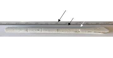 The bones which were heated for the purpose of this study were placed in the oven prior to it being switched on. This was done in a way as so to simulate a fire scenario whereby the temperature would not be uniform straight away. There was a notable difference in the form of those cut marks which were added pre- heat treatment compared with those added post- heat treatment. Figure 9 below shows a comparison between cut marks which were added both pre- and post- heat treatment.
Fig. 9. Radius with cut marks. Those marked with black arrows were placed pre- heat treatment. White arrow marked A was placed following 0.5 hours of heating. B placed following 1 hour and C placed following 1.5 hours
As can be seen in Figure 9, those cut marks which were placed post- heating experienced a greater degree of chipping compared with those added prior to heat treatment. The chipping was experienced as the cut was added. Extracting the blade from the bone generated bone fragments which were then burned off during the next heating session. This in turn produces a larger point of exit once exposed to heat compared to that of fresh bone. The trauma which was created by the knife leaves the bone more susceptible to processes such as cracking and fragmentation surrounding the area of the cut mark (de Gruchy and Rogers, 2002). This could extend from the cut mark itself or just be placed near to it as can be seen in Figures 3 and 9 respectively.
Following heat exposure, many of the cut marks placed on the bones remained visible with minimal detrimental alterations. It has been noted in multiple studies (Bertrand and Oxenham, 2015; Imaizumi, 2015; Pope and Smith, 2004; Shipman et al., 1984; Thompson, 2005; Ubelaker, 2009) that bones may experience shrinkage during heat treatment, however, this appeared to have little to no effect on the overall appearance of cut marks. The degree of the effect on the pre-existing trauma is often influenced by the temperature and duration of the fire (Bohnert et al., 1998). Shrinkage and deformation was not well documented within this study. With it still being in pilot stages, this is an area that further research should consider. Other studies into the effects of heat treatment on bone shrinkage have found that bone will reduce in volume throughout the burning process. However, this element proves difficult to quantify due to the difficulty in gaining precise volume measurements in cracked and fragmented bone (Imaizumi, 2015). A recent study by Harvig and Lynnerup (2013), looked into Computed Tomography (CT) as a way to take digital measurements for volume analysis of burnt bones where the bone may be presented in a more complex shape due to fragmentation. A further study in 2014 by Imaizumi et al., found that bone volume did not change until temperatures of 600°C. Once the temperature reached 700°C volume was drastically reduced, up until 1100°C by which time the volume had decreased by almost half. Based on this it can be suggested that volumes in this study would have showed little decrease since the temperature used was 600°C. Bone shrinkage occurs due to recrystallization of the hard matrix following the dehydration process. This leads to a contraction in the bones normal dimensions i.e. bone shrinkage.
Preliminary testing identified the need to document weight loss throughout the study, since alongside shrinkage, weight loss is one of the most significant factors brought about by heat treatment. The preliminary testing missed this variable and so this was rectified before the main study began. In this study the majority of bones experienced considerable weight loss which is highlighted in Table 2. Weight reduction in heat treated bones results from loss of water and organic materials including the main component, collagen Karlsbeek and Richter, 2006). A study by Karlsbeek and Richter, 2006 observed larger weight losses accompanied by a greater degree of shrinkage in cancellous bones compared with compact bone. This is thought to be because cancellous bone may contain more water than compact bone due to its more porous nature (Karlsbeek and Richter, 2006). It has been established that complete cremation of a human body has a difference in weight of approximately 1000g between males and females (Warren and Maples, 1997). A separate study corroborated this finding coming up with a similar figure seven years later (Bass and Jantz, 2004). Several studies into weight reduction in burnt bones have found a drastic decrease up until 400°C moving towards a plateau at around 700°C (Imaizumi at al., 2014; Karlsbeek and Richter, 2006; Hiller et al, 2003). Although within this study a consistent temperature of 600°C was used throughout each heating session, the bones were placed in the furnace from 0°C. Since the bones experienced a gentle increase of temperature followed by a period of cooling, this could have had an effect on the overall weight loss experienced. The process of continual heating and cooling could also have been a factor leading to the excessive heat fracturing which was experienced on many of the bones.
Looking at total cut mark length and the alterations it underwent following heat treatment, as can be seen in Table 3 there were no clear patterns of increase or decrease established. The majority of the tested bones experienced an overall decrease in total cut mark length, however, some showed an initial decrease followed by an increase towards the end of the heating sessions. Bone shrinkage and warping could have had an affect here; it is also possible that heat fracturing could have interfered with the cut marks leading to an inaccurate cut mark length being measured. Many of the fractures which were noted began in the area immediate surrounding a cut mark, with some cutting across existing marks. As aforementioned the trauma caused by the knife whilst placing a cut mark in bone may leave the bone more susceptible to fracturing and fragmentation at the site of the cut mark. The width of the cut marks was found to open up following continual heat exposure. However, this is based off of visual interpretations as no measurements were taken for cut mark width at the time of this study. This could have been due to alterations in the bone matrix occurring as a result of heat exposure.
The coloration within the cut marks themselves is also of interest. As can be seen in figure 9 there is a clear distinction between the coloration of the cut marks added pre- heat treatment to those added post- burning. Pope and Smith (2004) found that injuries pertaining to sharp force trauma which are accessible when burning occurs will exhibit accelerated colour changes compared to the exposed bone. Since the remaining soft tissue protecting the bone has been disturbed the stages of burning are able to proceed more completely and at a faster rate. There has been some debate surrounding the differences in heat-induced alterations between fleshed and de-fleshed bones (Kooi and Fairgrieve, 2013). Since all of the bones used in this study were semi-fleshed in nature it is difficult to determine the affects the remaining flesh would have had on the pre-existing trauma. Future work may wish to consider the differences in coloration of cut marks placed on both fleshed and defleshed bones following heat treatment.
It is difficult to replicate a fire scenario in a laboratory environment, therefore the aim of this study was to further recognise the general effects of heat on cut mark characteristics in bone. It is possible for heat-treatment to destroy all evidence of trauma as can be seen in the scapula depicted in Figure 10. However, this is not the case in all circumstances. Multiple studies have documented the survival of pre-existing trauma following heat exposure (De Gruchy and Rogers, 2002; Herrmann and Bennett, 1999; Kooi and Fairgrieve, 2013; Marciniak, 2009; Pope and Smith, 2004; Waltenberger and Schutkowski, 2017).
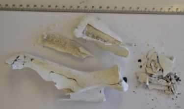
Fig. 10. A scapula showing excessive fragmentation and heat fracturing. The original cut mark which was placed on the bone has been completely destroyed during the heating process.
Kooi and Fairgrieve (2013) found the prevalence of recovered cut marks to be 55% and 81.8% for a smooth-edged knife and a serrated knife respectively. However, they also state that the predominance of recovered cut marks for the serrated edge knife may be due to the degree to which the samples were burnt as opposed to the fact they were produced by different knife types. The effect of heat treatment on cut marks produced by different knife types is an area which may require further research in the future. Other domestic tools such as handsaws, power saws and larger kitchen knives such as a cleaver will leave different characteristics on bone which will likely have their own individual reactions to heat treatment. This should be considered when applying this research to practical casework scenarios.
The knowledge gained during this study may be useful in the fields of both Forensic Anthropology and Forensic Science, alongside during any criminal investigations involving cremation and/or dismemberment. In terms of criminal investigations having a working knowledge of differences between pre- and post- cremation cut marks could allow links to be made between cases based upon this. Identifying a Modus Operandi (MO) that characteristically involves either pre- or post- heat treatment cut marks could in turn allow links to be made to individuals in order to aid in solving a potentially serious criminal case. Pre- heat treatment cut marks in bone could be an indication of some degree of torture, it may also help assess whether an individual was dismembered prior to, or following cremation.
Considerations
It should be noted that throughout the duration of the study only one individual performed all the bone incisions. Therefore, it is feasible to suggest that an individual of a different size with increased or decreased muscle mass may produce incisions with a different prevalence of cut mark traits. Since many of the characteristics which were analysed within this study required visual recognition, such as coloration, there is an element of subjectivity at this point. It is recommended that any professional body working in this field refer to other literature alongside this paper as a basis for evaluating colour and similar variables. Furthermore, their findings should undergo verification, especially in a live casework scenario.
It must be taken into consideration that animal bones were used throughout this study as an analogue for humans. Although there is considerable research stating that animal bones are a confirmed alternative for humans (Aerssens et al., 1998; Prat et al., 2012), there may be some difference in the way such bones react to burning. The animal bones used within this study were also of a juvenile nature. Juvenile bones are known to have greater elasticity and are able to withstand more mechanical stress before experiencing injury (Robbins et al., 2015). The elasticity of bone has been found to affect how completely a bone will burn. Juvenile bones are also less mineralized than those of an adult, this leads to greater shrinkage and heat-fracturing occurring following heat treatment (Bilesikian et al., 2002). Extensive fragmentation occurred for many of the bones analysed within this study. It is feasible to suggest that despite a high level of caution being undertaken, some of the smaller fragments of bone may have been lost between stages. This could have had an effect on the measured weight of each bone, however, only a slight difference would have been noted if any. Since the bones in this study were contained in a crucible it is less likely the loss of bone fragments would occur. In domestic fires where no attempt is made to isolate the bones from the surroundings such as during criminal activity, it is more likely that fragments of bone would be lost to the encompassing area.
Semi-fleshed bones were used throughout this study. It is likely that the presence of an increased level of soft tissue on the bones would impact upon any cut marks produced and analysed. The sawing action may be hindered by the presence of any excess soft tissue, however, once the knife had reached the bony material, it would be able to impart all cut mark characteristics as usual. The small amount of soft tissue present on the bones used in this study may have offered some protection from the heating process, although following 0.5 hours of heat treatment the remaining soft tissue had been burnt away leaving only the hard material exposed.
5. Conclusion
This research provides new observations into alterations of both pre- and post- heat treatment cut marks. It has previously been established that heat fracturing and fragmentation can have an effect on the appearance of cut marks on heat-treated bones, even acting to completely destroy them in certain circumstances. Pre- and post- burning cut marks have a difference in appearance, mainly in the coloration exhibited by them, and the level of chipping at the point of exit of the knife.
Previous literature suggested that bone shrinkage should have an effect on cut marks, however this study disclosed that the heating process did not have a significant effect on the form or deterioration of cut marks. Weight loss was experienced by all bones however this factor was independent of any cut mark characteristics and did not appear to have any effect in this area. Inconclusive results were gained surrounding the effect of heating on total cut mark length as the values from different bones did give any indication of a general pattern. Since this was only a pilot study it may be that there were not enough replicates to allow a pattern to be identified at this time.
There are various future directions which could be taken for any further research in this area. Looking into a range of knife types would allow information to be gained surrounding whether cut mark characteristics are more prominent, and therefore more likely to survive heat treatment, when produced by a certain knife type. This study focused mainly upon total cut mark length and any visual characteristics which experienced alterations. There are multiple other aspects of cut marks which can be looked into such as cut mark depth, slope angles, opening angle and floor angle amongst others. Noting whether differences in these variables occur in both pre- and post- heat treatment cut marks would allow the knowledge of this subject area to be further enhanced. A more extensive range of temperatures should be tested to determine whether an increased or decreased temperature has a similar effect or if there are notable differences in results. This study used a muffle furnace as the basis for the heat treatment however use of a more domestic fire could allow insight to be gained which would be better transferred to criminal investigations and the field of Forensic Science.
In terms of implications, this study suggests that within forensic and archaeological investigations, heat treatment of a bone will not necessarily act to obscure any preexisting trauma. Cut marks in burned bone can mostly be managed in a similar way to those on fresh bone. Although care must be taken when handling heat treated bones compared to fresh due to the fragility which occurs following heat fracturing and fragmentation.
Overall, this research provides new insights into cut marks on heat treated remains. New questions about heat alteration and pre- and post- burning cut marks have, however, been raised. Further research is required in order to aid in providing answers to these questions and provide added insight into this subject area.
Cite This Work
To export a reference to this article please select a referencing stye below:
Related Services
View all


Related Content
All TagsContent relating to: "Medicine"
The area of Medicine focuses on the healing of patients, including diagnosing and treating them, as well as the prevention of disease. Medicine is an essential science, looking to combat health issues and improve overall well-being.
Related Articles

DMCA / Removal Request
If you are the original writer of this dissertation and no longer wish to have your work published on the UKDiss.com website then please: